. 2011 Jun 15;22(12):2083-93.
doi: 10.1091/mbc.E10-11-0873. Epub 2011 Apr 27.

C4orf41 and TTC-15 are mammalian TRAPP components with a role at an early stage in ER-to-Golgi trafficking

Affiliations

C4orf41 and TTC-15 are mammalian TRAPP components with a role at an early stage in ER-to-Golgi trafficking

P James Scrivens et al. Mol Biol Cell. .

Abstract

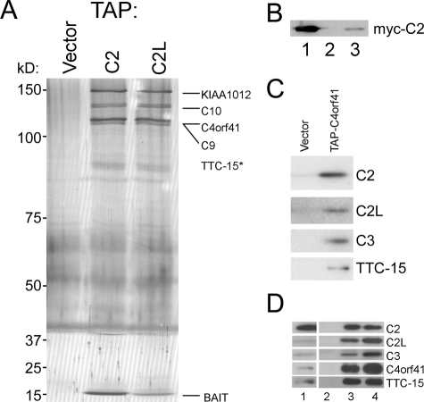
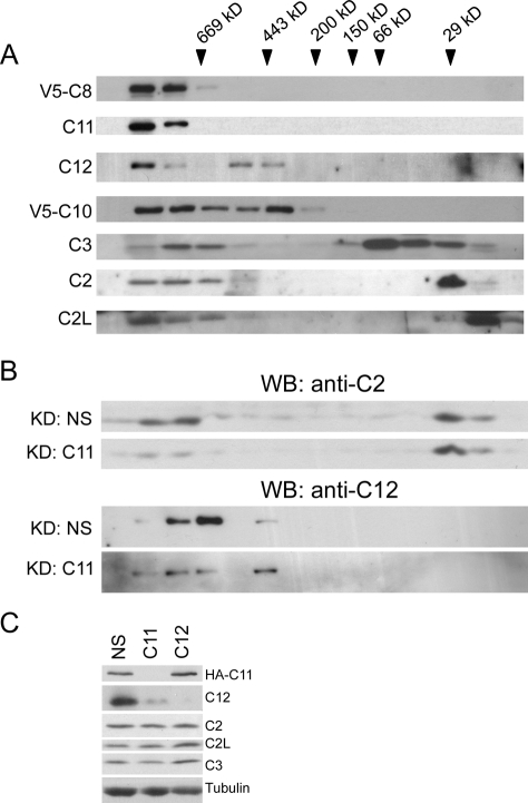
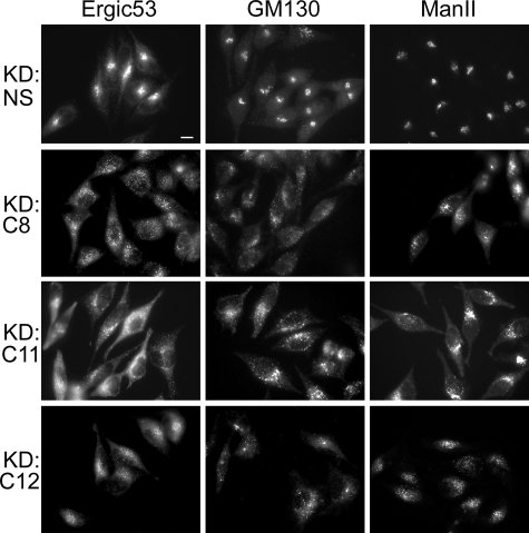
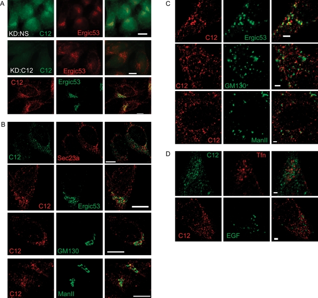
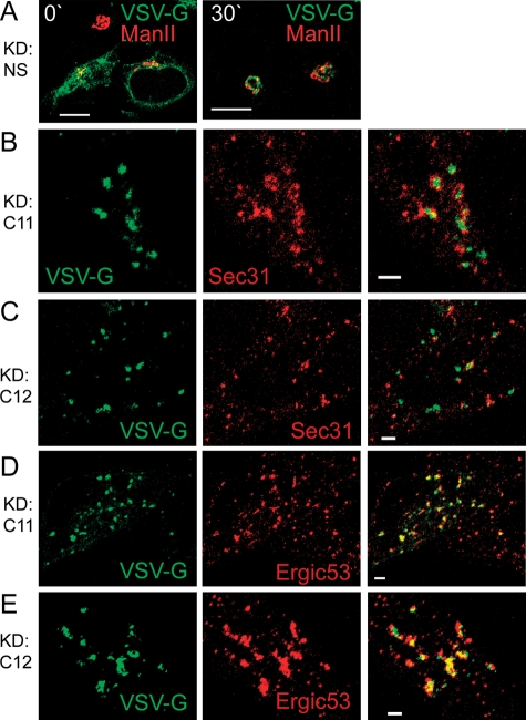
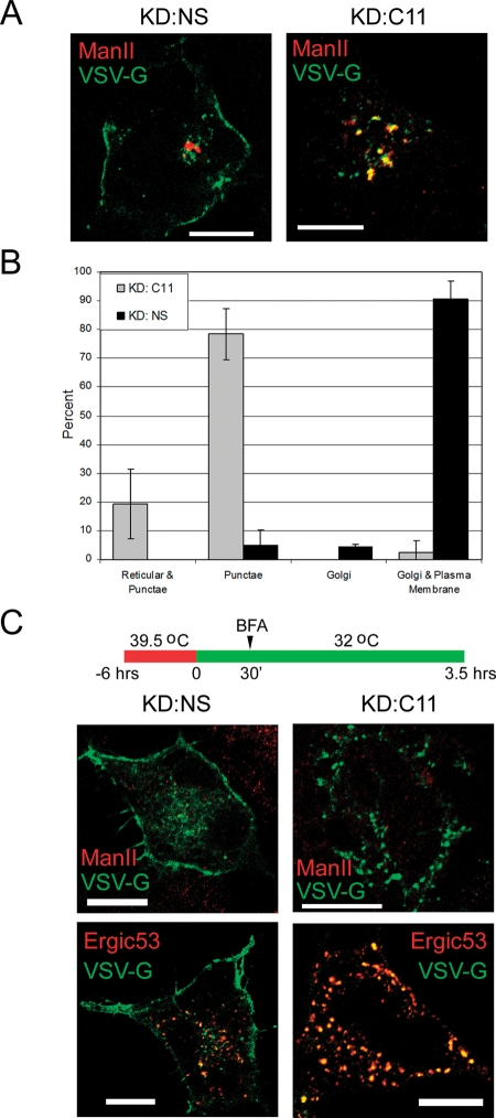
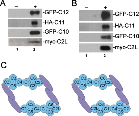
TRAPP is a multisubunit tethering complex implicated in multiple vesicle trafficking steps in Saccharomyces cerevisiae and conserved throughout eukarya, including humans. Here we confirm the role of TRAPPC2L as a stable component of mammalian TRAPP and report the identification of four novel components of the complex: C4orf41, TTC-15, KIAA1012, and Bet3L. Two of the components, KIAA1012 and Bet3L, are mammalian homologues of Trs85p and Bet3p, respectively. The remaining two novel TRAPP components, C4orf41 and TTC-15, have no homologues in S. cerevisiae. With this work, human homologues of all the S. cerevisiae TRAPP proteins, with the exception of the Saccharomycotina-specific subunit Trs65p, have now been reported. Through a multidisciplinary approach, we demonstrate that the novel proteins are bona fide components of human TRAPP and implicate C4orf41 and TTC-15 (which we call TRAPPC11 and TRAPPC12, respectively) in ER-to-Golgi trafficking at a very early stage. We further present a binary interaction map for all known mammalian TRAPP components and evidence that TRAPP oligomerizes. Our data are consistent with the absence of a TRAPP I-equivalent complex in mammalian cells, suggesting that the fundamental unit of mammalian TRAPP is distinct from that characterized in S. cerevisiae.


Figures

FIGURE 1:
FIGURE 1:
Identification of novel mammalian TRAPP components. (A) HEK293T cells were transfected with an empty vector (vector) or transfected with either TAP-C2 or TAP-C2L. Lysates were subjected to a two-step affinity purification and fractionated by SDS–PAGE. Bands were excised and subjected to mass spectrometric identification. In some cases, bands were not resolved and the entire eluate was analyzed by mass spectrometry. The bait band refers to either C2 (lane 2) or C2L (lane 3). TTC15/TRAPPC12 is marked with an asterisk since it was not identified in a gel slice but rather following mass spectrometric analysis of a non-gel-resolved protein preparation. Note that the uneven staining of the gel is due to the use of a discontinuous gradient in the resolving portion of the gel. (B) Lysates of HEK293T cells transfected with FLAG-C2L and myc-C2 (lane 1) were treated with preimmune rabbit serum (lane 2) or anti–FLAG IgG (lane 3), and the precipitates were subjected to Western analysis using anti–myc IgG. (C) Eluates following TAP purification were fractionated from cells transfected with an empty plasmid (vector) or with the TAP-C4orf41. The gel was transferred to a PVDF membrane and probed for the presence of the indicated TRAPP proteins. (D) HEK293T cells were transfected with HA-C4orf41. Lysates were incubated with preimmune serum (lane 2), anti-HA (lane 3), or anti-TTC15/C12 (lane 4). Immunoprecipitates were then fractionated by SDS–PAGE and probed for the presence of C2, C2L, C3, C12 or HA (indicating the presence of C4orf41/C11). Inputs representing 10% of the sample precipitated are shown in lane 1.
FIGURE 2:
FIGURE 2:
Size exclusion chromatography of TRAPP components. (A) Lysates from HEK293T cells left untransfected or transfected with V5-C8 or V5-C10 were fractionated on a Superdex 200 size exclusion column. Fractions (0.5 ml) were collected, and every second fraction was analyzed by Western analysis using antibodies against endogenous C2, C2L, C3, C11, and C12, or anti-V5 to detect transfected C8 or C10. (B) HeLa cells were treated with a nonspecific siRNA (KD: NS) or siRNA against C11 (KD: C11). Lysates were fractionated by size exclusion chromatography as above and subjected to Western analysis using anti–C2 antibody (top two panels) or anti–C12 antibody (bottom two panels). (C) HeLa cells transfected with HA-C11 were treated with a nonspecific siRNA (NS) or siRNA against C11 or C12. Equal amounts of the lysates were analyzed by Western blotting and probed for the presence of tubulin (loading control), C2, C2L, C3, C11 (using anti-HA), and C12.
FIGURE 3:
FIGURE 3:
Depletion of TRAPPC8, TRAPPC11, or TRAPPC12 results in Golgi dispersal. HeLa cells were treated with nonspecific siRNA or with siRNA against C8, C11, or C12 as indicated. Cells were stained with antibodies against ERGIC53, GM130, and mannosidase II, as indicated. The bar represents 10 μm. Quantitation of the phenotype using GM130 as a marker showed that 73% (n = 125), 88% (n = 43), and 88% (n = 42) of the cells from the C8, C11, and C12 knockdowns, respectively, displayed fragmented Golgi compared with 6% (n = 97), 2% (n = 52), and 2% (n = 52) from the nonspecific siRNA controls, respectively.
FIGURE 4:
FIGURE 4:
Localization of TRAPPC12. (A) HeLa cells were treated with nonspecific (NS; top row) or C12-specific (middle row) siRNAs and stained for C12 and ERGIC53, then visualized by epifluorescence microscopy (upper two rows). Maximum projection of confocal images from NS-treated cells (third row) recapitulates the punctate, perinuclear C12 localization seen by epifluorescence microscopy. (B) Single confocal slices of untreated HeLa cells costained for C12 and Sec23a, ERGIC53, GM130, or Man II. (C) HeLa cells were treated with 10 μM nocodazole for 1 h and then stained with antibodies against C12 and either ERGIC53, GM130, or Man II, as indicated. (D) HeLa cells were serum-starved for 2 h prior to incubation with fluor-tagged EGF or transferrin (Tfn). The scale bar in (A) and (B) represents 10 μm, whereas in (C) and (D) it represents 2 μm.
FIGURE 5:
FIGURE 5:
Depletion of either TRAPPC11 or TRAPPC12 perturbs ts045-VSV-G-GFP trafficking. HeLa cells were treated with nonspecific siRNA (A) or siRNA against either C11 (B and D) or C12 (C and E). After 48 h, cells were transfected with a plasmid encoding ts045-VSV-G-GFP. Cells were then shifted to 39.5°C for 6 h (A, left) and subsequently incubated at 32°C for 30 min (all other panels). Cells were then stained with anti-Sec31 or anti-ERGIC53, as indicated. The right panels in (B–E) represent merged images from the two panels directly to their left. The scale bar in (A) is 10 μm, and all other bars are 2 μm.
FIGURE 6:
FIGURE 6:
ts045-VSV-G-GFP is arrested in a BFA-resistant compartment upon TRAPPC11 depletion. (A) HeLa cells were treated as in Figure 5, except the cells were processed for fluorescence microscopy following 3.5 h at 32°C. Markers examined are indicated in each panel. (B) Quantification of the observed trafficking defect in C11-depleted HeLa cells. Approximately 30 cells per replicate were counted from 3 independent replicates following release from 39.5°C. (C) HeLa cells were treated as in (A), except 30 min into the 32°C incubation, BFA was added to the cultures. The scale bars represent 10 μm.
FIGURE 7:
FIGURE 7:
Mammalian TRAPP forms oligomers. (A) HEK293T cells were cotransfected with FLAG-C2L/myc-C2L, V5-C10/GFP-C10, V5-C11/HA-C11, or V5-C12/GFP-C12. Lysates were treated with preimmune rabbit serum (lane 1), anti–FLAG (for the C2L transfections), or anti–V5 (for the C10, C11, and C12 transfections) IgG and then probed with anti–myc (for the C2L transfection), anti–GFP (for the C10 and C12 transfections), or anti–HA (for the C11 transfection) IgG. (B) The lysates from (A) were fractionated by gel filtration chromatography. The high-molecular-weight TRAPP-containing fractions were pooled and immunoprecipitated and probed as in (A). (C) A model for the architecture of mammalian TRAPP built from yeast two-hybrid and coimmunoprecipitation data. Subunits shaded in blue are arranged based on the previously published architecture of the subcomplex (Kim et al., 2006). The high-molecular-weight subunits (C8–C12) are represented by a single mauve oval. Given interactions between high–molecular-weight components with proteins at both ends of the TRAPP “core,” a network of high-molecular-weight subunit interactions among themselves, and oligomerization of the complex as indicated in (A) and (B), two TRAPP “cores” could be bound via high-molecular-weight subunit interactions in trans. The two models differ with respect to the orientation of the second “core.” See the text for details.

References

    1. Allan BB, Moyer BD, Balch WE. Rab1 recruitment of p115 into a cis-SNARE complex: programming budding COPII vesicles for fusion. Science. 2000;289:444–448. - PubMed
    1. Bergmann JE, Singer SJ. Immunoelectron microscopic studies of the intracellular transport of the membrane glycoprotein (G) of vesicular stomatitis virus in infected Chinese hamster ovary cells. J Cell Biol. 1983;97:1777–1787. - PMC - PubMed
    1. Bonifacino JS, Glick BS. The mechanisms of vesicle budding and fusion. Cell. 2004;116:153–166. - PubMed
    1. Cai H, Yu S, Menon S, Cai Y, Lazarova D, Fu C, Reinisch K, Hay JC, Ferro-Novick S. TRAPPI tethers COPII vesicles by binding the coat subunit Sec23. Nature. 2007;445:941–944. - PubMed
    1. Cai H, Zhang Y, Pypaert M, Walker L, Ferro-Novick S. Mutants in trs120 disrupt traffic from the early endosome to the late Golgi. J Cell Biol. 2005;171:823–833. - PMC - PubMed

Publication types

MeSH terms

Substances