. 2011 Apr 27;31(17):6329-38.
doi: 10.1523/JNEUROSCI.5968-10.2011.

PSD-95 is required to sustain the molecular organization of the postsynaptic density

Affiliations

PSD-95 is required to sustain the molecular organization of the postsynaptic density

Xiaobing Chen et al. J Neurosci. .

Abstract

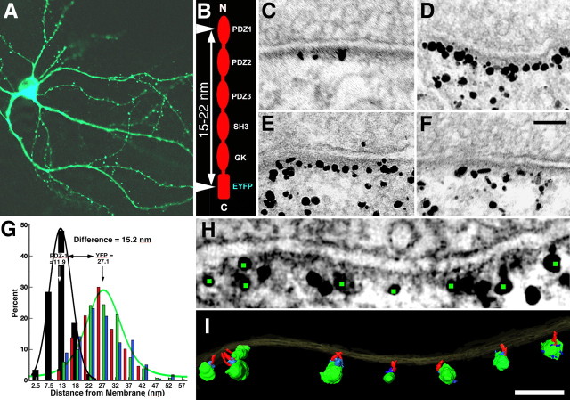
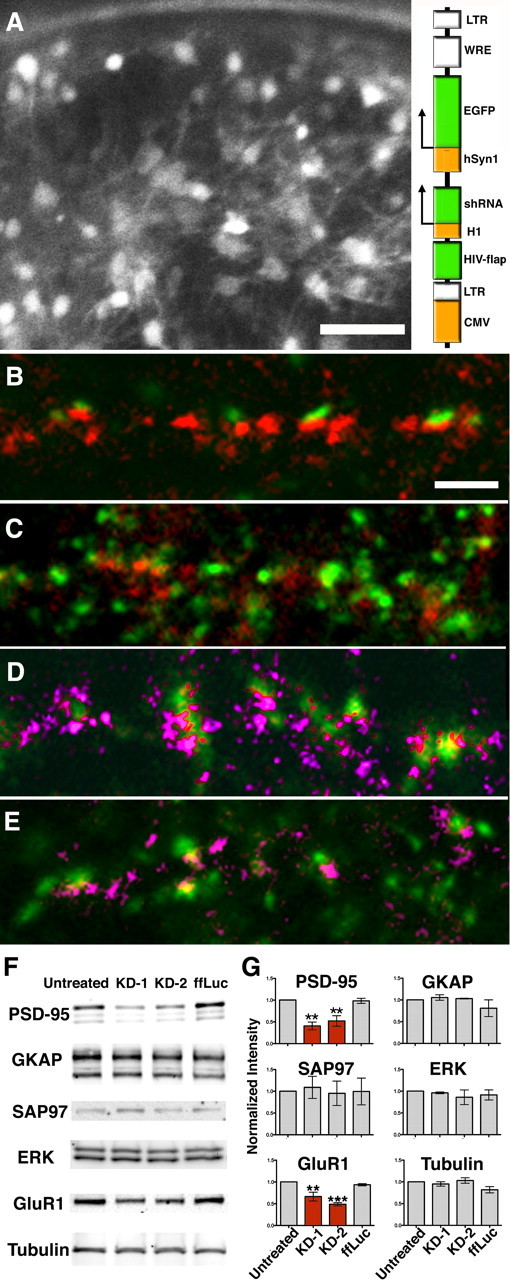
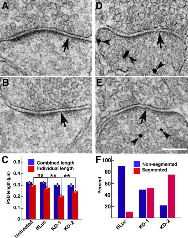
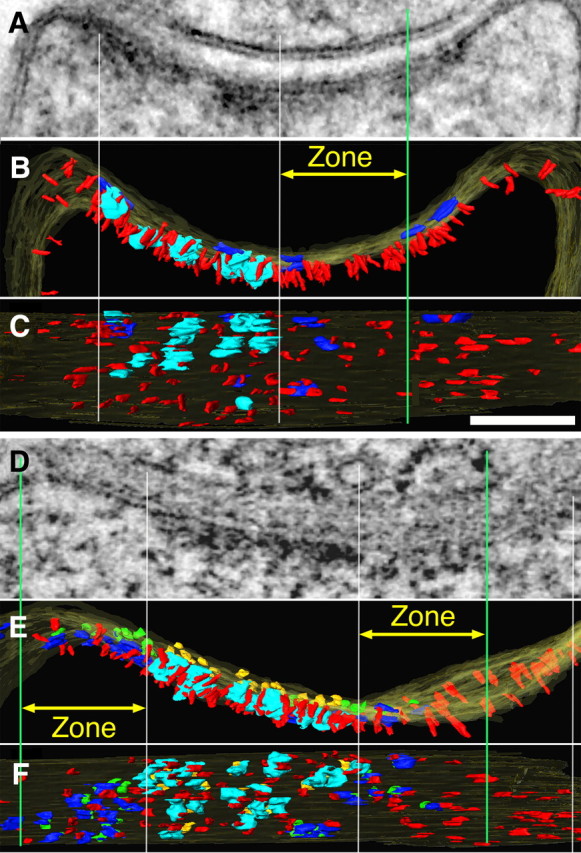
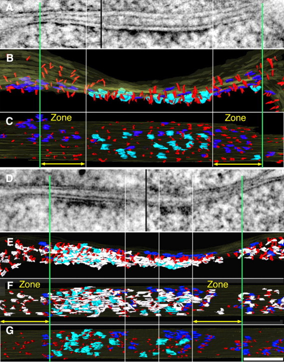
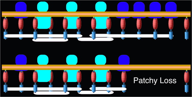
PSD-95, a membrane-associated guanylate kinase, is the major scaffolding protein in the excitatory postsynaptic density (PSD) and a potent regulator of synaptic strength. Here we show that PSD-95 is in an extended configuration and positioned into regular arrays of vertical filaments that contact both glutamate receptors and orthogonal horizontal elements layered deep inside the PSD in rat hippocampal spine synapses. RNA interference knockdown of PSD-95 leads to loss of entire patches of PSD material, and electron microscopy tomography shows that the patchy loss correlates with loss of PSD-95-containing vertical filaments, horizontal elements associated with the vertical filaments, and putative AMPA receptor-type, but not NMDA receptor-type, structures. These observations show that the orthogonal molecular scaffold constructed from PSD-95-containing vertical filaments and their associated horizontal elements is essential for sustaining the three-dimensional molecular organization of the PSD. Our findings provide a structural basis for understanding the functional role of PSD-95 at the PSD.


Figures

Figure 1.
Figure 1.
Conformation of PSD-95 in spine PSDs. A, Confocal image of 3 week hippocampal culture transfected with PSD-95-EYFP. B, Immunolabeled sites (arrowheads) on the tagged PSD-95 molecule showing predicted distance between these sites when PSD-95 is in extended configuration. C, Distribution of immunolabels using antibody to PDZ1 domain in 3 week hippocampal culture transfected with PSD-95-EYFP. D–F, Distributions of immunolabels with three different antibodies to EYFP site in parallel with 3 week hippocampal culture transfected with PSD-95-EYFP. G, Comparison of vertical separations of the immunolabels to the PDZ1 domain and to the EYFP site in the proximity of the PSD (<60 nm from the postsynaptic membrane). Each distribution (PDZ1, black; EYFPs red, green, blue) is fitted with a Gaussian (PDZ1, black; EYFP, green). H, Virtual section derived from the tomographic reconstruction of a spine synapse, immunolabeled with an antibody to the EYFP site. Green dots highlight centers of silver-enhanced immunogold particles associated with vertical filaments in the PSD (I). I, Surface rendering of the PSD in H. Vertical filaments ∼17 nm long (red) bridge immunogold particles and the postsynaptic membrane. Silver-enhanced Nanogold particles shown in green and structures thought to be parts of bridging antibodies shown in blue. For detailed measurements and statistics, see Results. Scale bars, 100 nm.
Figure 2.
Figure 2.
Knockdown of endogenous PSD-95 by RNAi. A, Many of the mature hippocampal neurons grown in a 3 mm gold chamber before high-pressure freezing are infected by the lentivirus, as indicated by the fluorescence from the shRNA reporter. Scale bar, 50 μm. Schematic representation of shRNA construct for generating lentivirus with a built-in EYFP reporter (inset, right). B, C, Immunofluorescence of cultures treated with shRNA to knock down PSD-95: ffLuc (control), scale bar 20 μm (B); KD-1 (C). Synaptophysin, green; and PSD-95, red. PSD-95 knockdown diminishes intensity of puncta labeled for PSD-95 (red) in C. D, E, Labeling for pan-MAGUKs (pink) shows that other MAGUKs are likely not affected by PSD-95 knockdown: untreated control (D); KD2 (E). F, Western blots for major PSD protein levels as follows: untreated, two PSD-95 knockdowns, and ffLuc control. G, Quantitative Western blots shows that only PSD-95 and GluR1 are significantly reduced by knockdown. Statistics results: one-way ANOVA, Dunnett test, PSD-95 (N = 3): untreated and KD1 (p = 0.002), untreated and KD2 (p = 0.007), untreated and ffLuc (p = 0.997); Tubulin (N = 3): untreated and KD1 (p = 0.866), untreated and KD2 (p = 0.95), untreated and ffLuc (p = 0.1); ERK (N = 3): untreated and KD1 (p = 0.67), untreated and KD2 (p = 0.98), untreated and ffLuc (p = 0.88); SAP97 (N = 3): untreated and KD1 (p = 0.99), untreated and KD2 (p = 0.998), untreated and ffLuc (p = 0.99); GKAP (N = 3): untreated and KD1 (p = 0.96), untreated and KD2 (p = 0.99), untreated and ffLuc (p = 0.45); GluR1 (N = 3): untreated and KD1 (p = 0.007), untreated and KD2 (p = 0.0005), untreated and ffLuc (p = 0.76). **p < 0.01, ***p < 0.001. CMV, Cytomegalovirus; LTR, long-term repeat; WRE, woodchuck regulatory element; H1, human H1 pol III promoter; HSyn1, human synapsin I pol II promoter; FLAP, HIV-1 flap element.
Figure 3.
Figure 3.
Patchy loss at the PSD correlates with PSD-95 knockdown. A, B, Conventional electron micrographs of spines from cultures with PSD-95 knocked down showing loss of patches of material from PSDs (arrows). C, Histograms comparing lengths of individual segments of the PSD (red) with the combined lengths of the PSD (blue) with and without PSD-95 knockdown. The highly significant difference (**p < 0.001, one-way ANOVA, Dunnett test; see Results) between average individual lengths is indicative of patchy loss. For detailed measurements and statistics, see Results. D, E, Spines affected by knockdown are independently identified by immunogold labeling for the EGFP reporter of shRNA (arrowheads). F, Correlation between PSD-95 knockdown identified by immunolabeling of shRNA reporter and patchy loss in control and knockdown experiments. Among the labeled spines, the percentage of total synapses showing either segmented (red bar) or nonsegmented (blue bar) PSD shown. There are fourfold to sixfold more segmented PSDs (red bars) after knockdown of PSD-95, a significant shift. Scale bar, 200 nm.
Figure 4.
Figure 4.
Knockdown of PSD-95 results in patchy loss of vertical filaments along with other components of the PSD. A, Thin section image through a synapse averaged from 31 virtual sections from the tomogram. B, C, Renderings of same tomogram in cross section (B) and en face view inside the spine (C) show that PSD-95 knockdown leads to reduction of vertical filaments in areas of patchy loss (between white center and green lines). PSD-95-containing vertical filaments, red; cytoplasmic side of NMDAR type structures, cyan; cytoplasmic side of AMPAR-type structures, dark blue. Vertical white lines align edges of PSD across the different views, green lines delineate zones for measurements (yellow arrows) of vertical filaments and AMPAR-type structures around the NMDAR-type structure cluster. D, Thin section image through another synapse averaged from 15 virtual sections from the tomogram. D–F, Renderings of the tomogram (D) in cross section (E) and en face viewed from inside the spine (F) with the additional rendering of extracellular side of NMDAR-type structures in gold and the extracellular side of AMPAR-type structures in green. Vertical filaments as well as AMPAR-type structures are depleted in an area of patchy loss. Scale bar, 100 nm. Measurements from renderings of tomograms: in C, zone right, 125 ×100 nm, number of vertical filaments 24, nearest-neighbor distances of vertical filaments 23 ± 11 nm (n = 85) and the number of AMPAR-type structures 3; in E, zones left and right, 125 ×100 nm, number of vertical filaments 19,20, nearest-neighbor distances of vertical filaments 30 ± 15 nm (n = 32), 28 ± 14 nm (n = 31), and the number of AMPAR-type structures 8,5.
Figure 5.
Figure 5.
A–G, Loss of core scaffold after PSD-95 knockdown: comparison of effects of PSD-95 knockdown (D–G) with a control synapse (A–C). A, Thin section image of control synapse is a montage of tomograms from two different projection angles (separated by black line) selected to maintain alignment of postsynaptic membrane. Surface renderings of same tomogram show the corresponding cross-section (B) and en face views from cytoplasmic side of the spine. C, Vertical PSD-95-containing filaments (red) form regular arrays covering the entire extent of the PSD. Vertical white lines align edges of NMDAR-type structures across the different views, while green lines delineate zones (yellow arrows) selected for measurements of vertical filaments and AMPAR-type structures around the central cluster, ∼170 nm in diameter, of NMDAR-type structures. The measurement zone is 125 nm wide and delimited by the section thickness, here 96 nm. Cytoplasmic side of NMDAR-type structures is shown in cyan, AMPAR-type structures are shown in dark blue. D–G, A PSD showing effects of PSD-95 knockdown. D, Thin section image of PSD-95 knockdown synapse illustrated as a montage of tomograms from two different projection angles (separated by black line) selected to maintain alignment of postsynaptic membrane. E, Rendered cross-section view of the PSD (D) showing vertical filaments, cytoplasmic side of major transmembrane structures, and horizontal elements associate with vertical filaments, color coded like control horizontal elements (white). E, En face view of the PSD facing spine cytoplasm. F, Complexes of horizontal filaments (white) associate with vertical filaments, data not shown in control. G, En face view of cytoplasmic side of the PSD without horizontal elements. Vertical white lines delineate zones on each side of the PSD showing patches of depletion of both vertical filaments (red) and horizontal elements (white), which appear as patchy loss in thin section images. Remaining dense material in PSD most closely reflects patches of NMDAR-type structures. In this example, the loss of NMDA receptor structures in the center of the PSD may reflect a perforation as the horizontal filaments are still present. Scale bar, 100 nm. Measurements from renderings of tomograms: in C, zone left and right, 125 × 96 nm, number of vertical filaments 29, 34; nearest-neighbor distances of vertical filaments 15 ± 5 nm (n = 41), 16 ± 6 nm (n = 45); and the number of AMPAR-type of structures 7,10; in F, zones left and right, 125 × 75 nm; number of vertical filaments 24, 20; nearest-neighbor distances of vertical filaments 31 ± 18 nm (n = 38), 30 ± 15 nm (n = 36) and the number of AMPAR-type structures 3,5.
Figure 6.
Figure 6.
Relationships between core scaffolding proteins and receptors in the PSD: control (above), PSD-95 knockdown (below). The N termini of regularly spaced vertical filaments of PSD-95 contact the postsynaptic membrane, and in some instances associate with AMPAR (blue) and NMDAR complexes (cyan). This arrangement imposes a layering of regularly spaced binding sites for other PSD-95 binding partners, thereby defining the molecular layering in the PSD. Horizontally oriented elements (white) intersect the C-terminal ends of the PSD-95 molecules, possibly cross-linking them. Knockdown of PSD-95 results in loss of patches of PSD-material, including both vertical and horizontal filaments as well as AMPAR structures, showing that both the core scaffold of the PSD and the organization of AMPAR complexes depend on PSD-95.

References

    1. Bats C, Groc L, Choquet D. The interaction between Stargazin and PSD-95 regulates AMPA receptor surface trafficking. Neuron. 2007;53:719–734. - PubMed
    1. Béïque JC, Lin DT, Kang MG, Aizawa H, Takamiya K, Huganir RL. Synapse-specific regulation of AMPA receptor function by PSD-95. Proc Natl Acad Sci U S A. 2006;103:19535–19540. - PMC - PubMed
    1. Bhattacharyya S, Biou V, Xu W, Schlüter O, Malenka RC. A critical role for PSD-95/AKAP interactions in endocytosis of synaptic AMPA receptors. Nat Neurosci. 2009;12:172–181. - PMC - PubMed
    1. Blanpied TA, Kerr JM, Ehlers MD. Structural plasticity with preserved topology in the postsynaptic protein network. Proc Natl Acad Sci U S A. 2008;105:12587–12592. - PMC - PubMed
    1. Chen L, Chetkovich DM, Petralia RS, Sweeney NT, Kawasaki Y, Wenthold RJ, Bredt DS, Nicoll RA. Stargazin regulates synaptic targeting of AMPA receptors by two distinct mechanisms. Nature. 2000;408:936–943. - PubMed

Publication types

MeSH terms

Substances

LinkOut - more resources