. 2011 May 15;214(Pt 10):1699-706.
doi: 10.1242/jeb.051581.

Cross-linking by protein oxidation in the rapidly setting gel-based glues of slugs

Affiliations

Cross-linking by protein oxidation in the rapidly setting gel-based glues of slugs

Andrew Bradshaw et al. J Exp Biol. .

Abstract

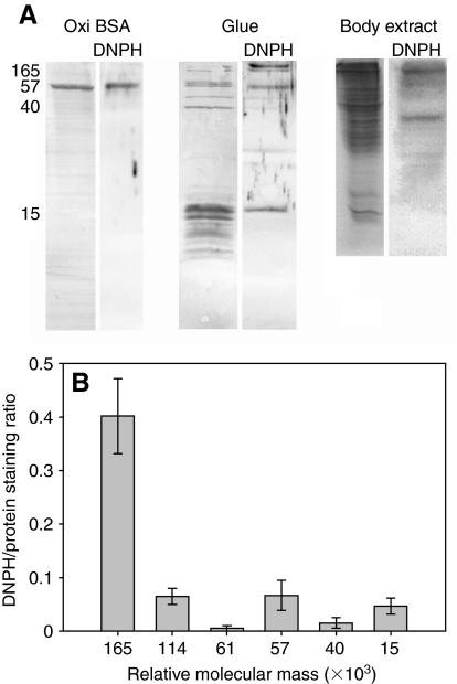
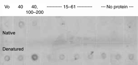
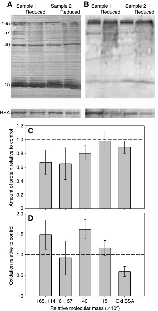
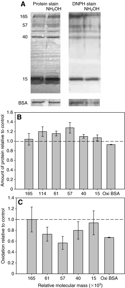
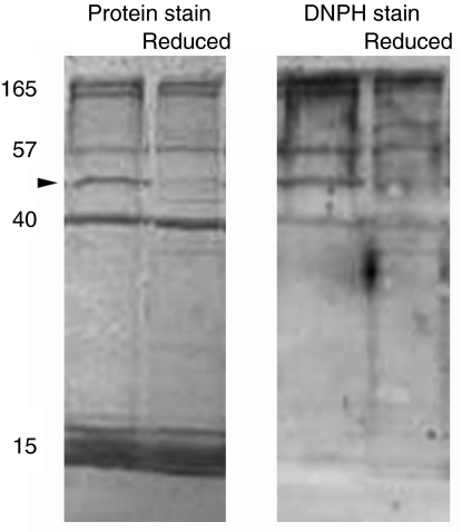
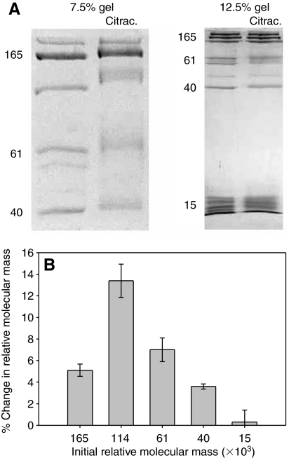
The terrestrial slug Arion subfuscus secretes a glue that is a dilute gel with remarkable adhesive and cohesive strength. The function of this glue depends on metals, raising the possibility that metal-catalyzed oxidation plays a role. The extent and time course of protein oxidation was measured by immunoblotting to detect the resulting carbonyl groups. Several proteins, particularly one with a relative molecular mass (M(r)) of 165 x 10³, were heavily oxidized. Of the proteins known to distinguish the glue from non-adhesive mucus, only specific size variants were oxidized. The oxidation appears to occur within the first few seconds of secretion. Although carbonyls were detected by 2,4-dinitrophenylhydrazine (DNPH) in denatured proteins, they were not easily detected in the native state. The presence of reversible cross-links derived from carbonyls was tested for by treatment with sodium borohydride, which would reduce uncross-linked carbonyls to alcohols, but stabilize imine bonds formed by carbonyls and thus lead to less soluble complexes. Consistent with imine bond formation, sodium borohydride led to a 20-35% decrease in the amount of soluble protein with a M(r) of 40-165 (x 10³) without changing the carbonyl content per protein. In contrast, the nucleophile hydroxylamine, which would competitively disrupt imine bonds, increased protein solubility in the glue. Finally, the primary amine groups on a protein with a M(r) of 15 x 10³ were not accessible to acid anhydrides. The results suggest that cross-links between aldehydes and primary amines contribute to the cohesive strength of the glue.


Figures

Fig. 1.
Fig. 1.
The extent of oxidation of the proteins in Arion subfuscus glue, based on anti-DNPH immuno-staining for carbonyl groups. (A) Sample of oxidized BSA (positive control), glue and whole body extract. Left lanes show each blot stained for total protein content using Coomassie Blue R-250. Right lanes (DNPH) show the corresponding anti-DNPH immune blots. The numbers on the left show relative molecular mass (×103). (B) Quantification of the extent of oxidation of selected proteins in the glue based on signal intensity. Values are the ratios of staining intensity of the immunostain relative to the staining intensity of Coomassie Blue for that protein on a duplicate blot (mean ± s.e.m., N=6). Oxidized BSA (positive control) had a DNPH/protein staining ratio of 0.37±0.04.
Fig. 2.
Fig. 2.
DNPH immunostained dot blot of proteins separated by gel filtration. The top row shows proteins in their native state, while the bottom row shows the same samples denatured before adding to the membrane. The numbers across the top show the relative molecular mass (×103) of the proteins in each fraction. The first spot contains the void volume fraction (Vo), which may contain giant polysaccharides and some protein. The second spot contains asmp-40, which elutes as a giant complex near the void volume. The third spot contains primarily asmp-114 and asmp-165, with a small amount of asmp-40. The fourth through seventh spots contain asmp-15, asmp-57 and asmp-61, with the proteins with a Mr of ∼15×103 making up the bulk of the material. The remaining spots contained no proteins that were visible on SDS-PAGE, though there was some material, as indicated by a yellow color in the samples.
Fig. 3.
Fig. 3.
The effect of sodium borohydride on the proteins in A. subfuscus glue. (A) Coomassie-Blue-stained blot showing the proteins present in the glue, and (B) anti-DNPH immunostained duplicate blot. Two different samples are shown; for each, the left lane is untreated whereas the right lane was reduced with borohydride. Oxidized BSA controls treated in the same way are shown at the bottom. The numbers on the left show relative molecular mass (×103). (C) Amount of soluble protein in borohydride-treated samples relative to untreated controls for selected proteins. (D) Extent of oxidation of selected proteins from borohydride-treated samples relative to their untreated counterpart. Some proteins were grouped together because they were difficult to quantify separately. The dashed lines in C and D correspond to the y-value if there was no difference between the control and treatment. Values are means ± s.e.m. (N=5).
Fig. 4.
Fig. 4.
The effect of hydroxylamine on the proteins in A. subfuscus glue. (A) The left two lanes show the Coomassie-Blue-stained blot, and the right two lanes show the DNPH immunostained duplicate blot. For each pair, the left lane is untreated and the right lane is treated with hydroxylamine (NH2OH). Oxidized BSA controls treated in the same way are shown at the bottom. The numbers on the left show relative molecular mass (×103). (B) Amount of soluble protein in hydroxylamine-treated samples relative to untreated controls for selected proteins. (C) Extent of oxidation of selected proteins from hydroxylamine-treated samples relative to their untreated counterpart. Note that asmp-114 is not included in this graph because its peak could not be easily distinguished from the strong signal for asmp-165. The dashed lines in B and C correspond to the y-value if there was no difference between the control and treatment. Values are means ± s.e.m. (N=5).
Fig. 5.
Fig. 5.
Analysis of A. subfuscus glue that was frozen before setting. The left two lanes show the Coomassie-Blue-stained blot, and the right two lanes show the DNPH immunostained duplicate blot. For each pair, the left side is the control and the right side is reduced with NaBH4. In this procedure, the control and reduced samples are not from the same animal. The arrowhead indicates asmp-44, which is prominent in immediately frozen samples. The numbers on the left show relative molecular mass (×103).
Fig. 6.
Fig. 6.
The effect of citraconylation at pH 6 on the relative molecular mass of the proteins in A. subfuscus glue. (A) Coomassie-Blue-stained SDS-PAGE. A low- and a high-percentage gel are shown (7.5 and 12.5%, respectively). For each pair, the left lane is the control and the right lane is treated with citraconic anhydride (citrac.) to modify primary amines. The numbers on the left of each show relative molecular mass (×103). (B) Percent change in relative molecular mass of selected proteins after citraconylation (mean ± s.e.m., N=3).

References

    1. Berlett B. S., Stadtman E. R. (1997). Protein oxidation in aging, disease, and oxidative stress. J. Biol. Chem. 272, 20313-20316 - PubMed
    1. Broomell C. C., Mattoni M. A., Zok F. W., Waite J. H. (2006). Critical role of zinc in hardening of Nereis jaws. J. Exp. Biol. 209, 3219-3225 - PMC - PubMed
    1. Chen T., Embree H. D., Brown E. M., Taylor M. M., Payne G. F. (2003). Enzyme-catalyzed gel formation of gelatin and chitosan: potential for in situ applications. Biomaterials 24, 2831-2841 - PubMed
    1. Coyne K. J., Qin X. X., Waite J. H. (1997). Extensible collagen in mussel byssus: a natural block copolymer. Science 277, 1830-1832 - PubMed
    1. Dickinson G. H., Vega I. E., Wahl K. J., Orihuela B., Beyley V., Rodriquez E. N., Everett R. K., Bonaventura J., Rittschof D. (2009). Barnacle cement: a polymerization model based on evolutionary concepts. J. Exp. Biol. 212, 3499-3510 - PMC - PubMed

Publication types