. 2011 Jul;85(13):6736-49.
doi: 10.1128/JVI.00233-11. Epub 2011 Apr 27.

Delayed cytosolic exposure of Japanese encephalitis virus double-stranded RNA impedes interferon activation and enhances viral dissemination in porcine cells

Affiliations

Delayed cytosolic exposure of Japanese encephalitis virus double-stranded RNA impedes interferon activation and enhances viral dissemination in porcine cells

Lyre Anni Espada-Murao et al. J Virol. 2011 Jul.

Abstract

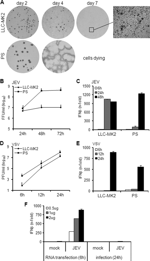
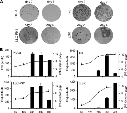
Interferon is a principal component of the host antiviral defense system. In this study, abortive focus formation by Japanese encephalitis virus (JEV) in primate cells was accompanied by early interferon induction, while productive focus formation in porcine cells was associated with a late interferon response. Neutralization antibodies against interferon relieved the restricted infection in primate cells, and increasingly larger foci were generated as treatment with exogenous interferon was delayed, thereby establishing a solid correlation between interferon response and viral dissemination. However, delayed interferon induction in JEV-infected porcine cells occurred in the absence of active inhibition by the virus. We further demonstrated that JEV mediates interferon activation through double-stranded RNA and cytosolic pattern recognition receptors. Immunofluorescence and subcellular fractionation studies revealed that double-stranded RNA is concealed in intracellular membranes at an early phase of infection but eventually appears in the cytosol at later periods, which could then allow detection by cytosolic pattern recognition receptors. Interestingly, cytosolic exposure of double-stranded RNA was delayed in porcine cells compared to primate cells, independent of total double-stranded RNA levels and in correlation with the timing of the interferon response. Furthermore, when double-stranded RNA was artificially introduced into the cytosol of porcine cells, more rapid and robust interferon activation was triggered than in viral infection. Thus, cytosolic exposure of JEV double-stranded RNA is imperative for interferon induction, but in cell lines (e.g., porcine cells) with delayed emergence of cytosolic double-stranded RNA, the interferon response is late and viral dissemination is consequently enhanced.


Figures

Fig. 1.
Fig. 1.
Focus formation, growth kinetics, and IFN activation by JEV in rhesus and porcine cells. (A) LLC-MK2 and PS cells were infected with JEV, and foci were detected by immunostaining them on the indicated days postinfection. The inset was obtained using ×100 magnification. (B) Viral titer of supernatants from JEV-infected cells (MOI, 1) at the indicated hours postinfection. The error bars indicate standard deviations of the means. (C) IFN-β mRNA expression in cells infected with JEV at an MOI of 1 was quantified via real-time quantitative RT-PCR. The results are expressed as fold increase over mock-treated cells and normalized to GAPDH and β-actin for LLC-MK2 and PS cells, respectively. The error bars indicate standard deviations of the means. (D) Viral titer of supernatants from VSV-infected cells (MOI, 0.1) at the indicated hours postinfection. The error bars indicate standard deviations of the means. (E) IFN-β mRNA expression in cells infected with VSV at an MOI of 0.1 was quantified via real-time quantitative RT-PCR. The results for IFN-β mRNA are expressed as the fold increase over mock-treated cells and normalized to GAPDH and β-actin for LLC-MK2 and PS, respectively. (F) Total RNA from mock-treated or JEV-infected LC-MK2 cells was transfected for 6 h in PS cells at 0.5, 1, or 2 μg. Another set of cells was mock treated or infected with JEV at an MOI of 1 for 24 h. IFN activation was measured by real-time quantitative RT-PCR. The results for IFN-β mRNA are expressed as the fold increase over mock-treated cells and normalized to β-actin. The error bars indicate standard deviations of the means.
Fig. 2.
Fig. 2.
Antiviral activity of JEV supernatants and roles of IFN and viral proteins. Supernatants (supe) from mock-treated or JEV-infected cells were UV inactivated and tested for the ability to block focus formation. JEV-infected cells were treated with the supernatants immediately after the viral adsorption period, and focus staining was performed 2 days postinfection. (A) LLC-MK2 supernatants harvested at the indicated times were applied on infected Vero cells, while PS supernatants were applied on infected PS cells. (B) Supernatant from JEV-infected LLC-MK2 cells was preincubated with 0, 0.05, and 0.5 μg/ml anti-human IFN-β prior to application on infected Vero cells. Mock supernatant was used as a control. (C) LLC-MK2 supernatants were applied on infected LLC-MK2 or PS cells.
Fig. 3.
Fig. 3.
Focus formation and IFN activation in other cell lines. (A) HeLa, LLC-PK1, PK, and ESK cells were infected with JEV, and foci were detected by immunostaining at the indicated days postinfection. (B) IFN activation (bar graph) and viral RNA titers (line graph) were measured in JEV-infected cells (MOI, 1) at the indicated times postinfection by real-time quantitative RT-PCR. The results for IFN-β mRNA are expressed as the fold increase over mock-treated cells. The results for JEV RNA are expressed as log10 copies of RNA per 1 μg total RNA. The values were normalized to GAPDH and β-actin for primate and porcine cells, respectively. The error bars indicate standard deviations of the means.
Fig. 4.
Fig. 4.
Regulation of focus formation by IFN and timing of the IFN response. (A) JEV focus formation was assessed at 3 and 5 days (d) p.i. in LLC-MK2 and HeLa cells treated with an anti-IFN antibody cocktail or a control IgG/serum cocktail. (B and C) Vero cells were infected with JEV and then treated with recombinant human IFN-β at the indicated times postinfection. Focus staining was performed 3 days postinfection. (B) Cells were treated with 0 U IFN-β at 1.5 h postinfection or 1,000 U IFN-β at 1.5, 12, 24, 36, and 48 h postinfection. (C) Cells were treated with 0, 10, 100, and 1,000 U IFN-β at 12 and 24 h postinfection.
Fig. 5.
Fig. 5.
Effect of JEV on the IFN activation pathway. (A) LLC-MK2 or PS cells were mock treated (m) or infected with JEV at an MOI of 1 for 24 h. Cellular extracts were subjected to immunoblotting for the proteins indicated on the left, with β-actin as the internal control. (B and C) PS cells were mock treated or infected with JEV at an MOI of 1 for 18 h, and then the indicated amounts of total RNA from mock-treated or JEV-infected LLC-MK2 cells were transfected for another 6 h. IFN activation (B) and viral RNA titers (C) were measured by real-time quantitative RT-PCR. The results for IFN-β mRNA are expressed as the fold increase over mock-treated cells. The results for JEV RNA are expressed as the log10 number of RNA copies per 1 μg total RNA. The values were normalized to β-actin, and the error bars indicate standard deviations of the means.
Fig. 6.
Fig. 6.
Role of double-stranded RNA in IFN activation and quantitation of intracellular viral RNA and double-stranded RNA. (A and B) Five hundred nanograms of total RNA from mock-treated or JEV-infected cells was incubated with 1 U RNase III, 1 U RNase R, or glycerol (no enzyme). (A) Pretreated RNA was transfected in HeLa cells for 6 h, and IFN activation was measured by real-time quantitative RT-PCR. The results are expressed as the fold increase over cells transfected with mock RNA and normalized to GAPDH. The error bars indicate standard deviations of the means. (B) A similar set of pretreated RNAs was subjected to JEV RNA quantitation by real-time RT-PCR. The results are expressed as the log10 number of RNA copies per 1 μg total RNA and normalized to GAPDH. The error bars indicate standard deviations of the means. (C and E) LLC-MK2 or PS cells were infected with JEV at an MOI of 1 (C) or PS cells were infected at MOIs of 1, 5, and 10 (E). IFN activation (bar graphs) and viral RNA titers (line graphs) were measured at the indicated times postinfection by real-time quantitative RT-PCR. The results for IFN-β mRNA are expressed as the fold increase over mock-treated cells. The results for JEV RNA are expressed as the log10 copies of RNA per 1 μg total RNA. The values were normalized to GAPDH and β-actin for LLC-MK2 and PS cells, respectively. The error bars indicate standard deviations of the means. (D) Immunodetection of dsRNA in LLC-MK2 or PS cells mock treated or infected with JEV at the indicated MOIs 24 h postinfection. The rate of dsRNA expression was obtained by calculating the mean percentage of positively stained cells in five fields. The values at the top right indicate the rates of dsRNA expression ± standard deviations. (F) Quantitation of double-stranded RNA via a solid-phase immunosorbent method. Hot-phenol-extracted total RNA (1 μg) from uninfected or JEV-infected cells was incubated with anti-dsRNA antibody (Ab) or nonspecific antibody on immunosorbent plates. Antibody-bound RNA was harvested via proteinase K/SDS treatment, reextracted, and quantified by real-time RT-PCR using primers for JEV. Untreated RNA was also quantified as the input viral RNA. The results represent the log10 copies of viral RNA. The error bars indicate standard deviations of the means.
Fig. 7.
Fig. 7.
Subcellular localization of double-stranded RNA. (A) Hot-phenol-extracted RNA from intracellular membrane fractions of mock-treated or JEV-infected LLC-MK2 cells was incubated with nonspecific or dsRNA-specific antibody on immunosorbent plates or left untreated (input). Antibody-bound RNA was harvested by proteinase K/SDS treatment, reextracted, and quantified by real-time RT-PCR using JEV-specific primers. The results are expressed as log10 copies of viral RNA. The error bars indicate standard deviations of the means. (B and C) Membrane-specific permeabilization of LLC-MK2 cells using NP-40 to permeabilize all membranes or SLO to permeabilize only the plasma membrane. Nuclear staining was achieved using DAPI. (B) Immunodetection of IRF-3 or calregulin in NP-40- or SLO-permeabilized LLC-MK2 cells. (C) Immunodetection of dsRNA and E protein in NP-40- or SLO-permeabilized LLC-MK2 cells at various times postinfection by JEV (MOI, 1). (D) Colocalization of dsRNA and calregulin in LLC-MK2 cells mock treated or infected with JEV (MOI, 1) 24 h postinfection. Nuclear staining was achieved using DAPI.
Fig. 8.
Fig. 8.
Quantitation of cytosolic viral RNA and double-stranded RNA. (A and B) LLC-MK2 or PS cells were infected with JEV (MOI, 1), and cytosolic fractions were obtained at various times postinfection. (A) RNA was isolated from cytosolic extracts using the spin column method and subjected to viral RNA quantification by real-time RT-PCR. The results are expressed as log10 copies of viral RNA per 1 × 10⁶ cells. The error bars indicate standard deviations of the means. The asterisks indicate the statistical significance (P < 0.001) between the comparison groups. (B) Phenol-extracted RNA from the cytosolic extract was incubated with nonspecific or dsRNA-specific antibody on immunosorbent plates or left untreated (input). Antibody-bound RNA was harvested by proteinase K/SDS treatment, reextracted, and quantified by real-time RT-PCR using JEV-specific primers. The results are expressed as log10 copies of viral RNA per 100 ng total RNA. The error bars indicate standard deviations of the means. The asterisks indicate the statistical significance (P < 0.001) between the comparison groups. (C) Immunodetection of dsRNA and E protein in NP-40- or SLO-permeabilized cells at various times postinfection by JEV (MOI, 1). NP-40 was used to permeabilize all membranes or SLO to permeabilize only the plasma membrane. Nuclear staining was achieved using DAPI. The white arrowheads indicate cells with intense dsRNA staining.

References

    1. Baum A., García-Sastre A. 2010. Induction of type I interferon by RNA viruses: cellular receptors and their substrates. Amino Acids 38:1283–1299 - PMC - PubMed
    1. Chang T. H., Liao C. L., Lin Y. L. 2006. Flavivirus induces interferon-beta gene expression through a pathway involving RIG-I dependent IRF-3 and PI3K-dependent NF-κB activation. Microb. Infect. 8:157–171 - PubMed
    1. Chen Z., et al. 2007. GB virus B disrupts RIG-I signaling by NS3/4A-mediated cleavage of the adaptor protein MAVS. J. Virol. 81:964–976 - PMC - PubMed
    1. Diamond M. S., et al. 2000. Modulation of dengue virus infection in human cells by alpha, beta and gamma interferons. J. Virol. 74:4957–4966 - PMC - PubMed
    1. Fredericksen B. L., Smith M., Katze M. G., Shi P. Y., Gale M., Jr 2004. The host response to West Nile Virus infection limits viral spread through the activation of the interferon regulatory factor 3 pathway. J. Virol. 78:7737–7747 - PMC - PubMed

Publication types

LinkOut - more resources