. 2011 Jul;85(13):6117-26.
doi: 10.1128/JVI.00378-11. Epub 2011 Apr 27.

RAB11A is essential for transport of the influenza virus genome to the plasma membrane

Affiliations

RAB11A is essential for transport of the influenza virus genome to the plasma membrane

Amie J Eisfeld et al. J Virol. 2011 Jul.

Abstract

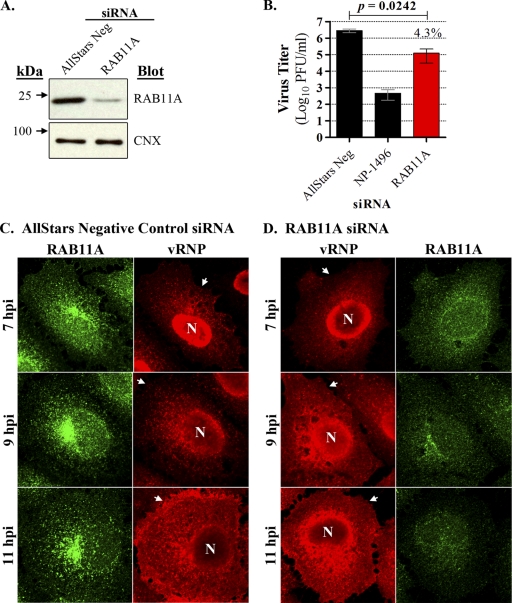
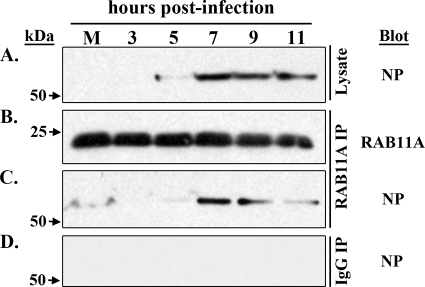
Influenza A virus assembly is a complex process that requires the intersection of pathways involved in transporting viral glycoproteins, the matrix protein, and viral genomes, incorporated in the viral ribonucleoprotein (vRNP) complex, to plasma membrane sites of virion formation. Among these virion components, the mechanism of vRNP delivery is the most incompletely understood. Here, we reveal a functional relationship between the cellular Rab11 GTPase isoform, RAB11A, and vRNPs and show that RAB11A is indispensable for proper vRNP transport to the plasma membrane. Using an immunofluorescence-based assay with a monoclonal antibody that recognizes nucleoprotein in the form of vRNP, we demonstrate association between RAB11A and vRNPs at all stages of vRNP cytoplasmic transport. Abrogation of RAB11A expression through small interfering RNA (siRNA) treatment or disruption of RAB11A function by overexpression of dominant negative or constitutively active proteins caused aberrant vRNP intracellular accumulation, retention in the perinuclear region, and lack of accumulation at the plasma membrane. Complex formation between RAB11A and vRNPs was further established biochemically. Our results uncover a critical host factor with an essential contribution to influenza virus genome delivery and reveal a potential role for RAB11A in the transport of ribonucleoprotein cargo.


Figures

Fig. 1.
Fig. 1.
Monoclonal antibody 3/1 recognizes vRNP in the cytoplasm of influenza virus-infected cells. MDCK cells grown on coverslips were infected with WSN virus (MOI of 3) and fixed at 3, 5, and 7 hpi before being subjected to coimmunofluorescence and FISH staining. MAb 3/1, which recognizes influenza virus NP, is indicated in red, and the PB2 negative-sense vRNA is shown in green. (A) Individual staining profiles are shown for NP and vRNA, together with a merged image, for each time point. Merged images show Hoechst nuclear stains. (B) Enlarged portions of the boxed sections in merged frames of panel A, highlighting colocalization of vRNA and NP.
Fig. 2.
Fig. 2.
Cytoplasmic vRNPs colocalize with RAB11A during transport from the MTOC to the plasma membrane. A549 cells grown on coverslips were mock infected or infected with WSN virus and stained with MAb 3/1 (vRNP; red) and a polyclonal rabbit antibody against RAB11A (green). Mock-infected cells were counter-stained with Hoechst 33258 to identify nuclei (blue). Representative spatiotemporal distribution patterns are shown for each staining condition at each time point. For each panel, frame i depicts merged images of vRNP, RAB11A, and Hoechst-stained nuclei, as indicated. Arrows point to the plasma membrane boundaries; N indicates the nucleus. Individual vRNP and RAB11A staining profiles are shown in frames ii and iii, respectively. Enlarged (3×) images of individual and merged vRNP and RAB11A distributions from the boxed regions in frames i are shown in the in frames iv for vRNP, v for RAB11A, and vi for merged images. The time point is indicated at the top of each panel, and a staining key is shown in the box in the bottom right corner. To facilitate observation of the details of vRNP and RAB11A localization, original images were acquired with a 40× objective and a 3× zoom. All images are representative of several independent experiments.
Fig. 3.
Fig. 3.
RAB11A knockdown impairs vRNP transport in influenza virus-infected cells. (A) A549 cells were transfected with nontargeting siRNA (AllStars Neg) or siRNA targeting RAB11A, and cell lysates were subjected to immunoblot analysis 48 h after transfection. siRNA treatments are indicated at the top, molecular mass standards are shown to the left, and detected proteins are indicated to the right of each panel. (B) Parallel A549 cultures were transfected with AllStars Neg siRNA, siRNA targeting influenza virus NP mRNA (NP-1496), or siRNA targeting RAB11A, and after 48 h cultures were directly inoculated with WSN virus. Supernatants were harvested at 48 h postinfection, and titers were determined by plaque assay in MDCK cells. Titers for each condition are represented by an average of triplicate infections, and variation is indicated by standard deviation. A paired Student's t test was performed to compare the AllStars Neg siRNA treatment with that of RAB11A siRNA, and the resultant P value is indicated at the top. The percentage of replication observed in RAB11A siRNA-transfected cells relative to that in cells transfected with the AllStars Neg control is indicated above the RAB11A bar. (C and D) A549 cells grown on coverslips were transfected with AllStars Neg siRNA or siRNA targeting RAB11A and then infected with WSN virus for vRNP trafficking analysis. Panel C shows the vRNP (red) and RAB11A (green) staining profiles for nontargeting siRNA while panel D shows the same for siRNA targeting RAB11A. Time points are indicated to the left. Arrows indicate the plasma membrane boundaries. Data are representative of two independent experiments. N, nucleus.
Fig. 4.
Fig. 4.
RAB11A mutant proteins disrupt vRNP trafficking during the late-stage of influenza virus infection. (A) RAB11A GFP fusion protein behavior in plasmid-transfected, uninfected cells. The upper left portion of this panel shows immunoblots of the GFP fusion proteins. The specific overexpressed protein is indicated above each lane and molecular mass markers (kDa) are indicated to the left. Images show the localization patterns for GFP alone, GFP-RAB11A-Q70L (constitutively active), and GFP-RAB11A-S25N (dominant negative), as indicated on the figure, in A549 cells. Cells were counterstained with Hoechst 33258. (B to D) vRNP distribution in WSN virus-infected A549 cells expressing RAB11A GFP fusion proteins. A549 cells were transfected with plasmids expressing GFP alone (B), GFP-RAB11A-Q70L (C), or GFP-RAB11A-S25N (D) and were subsequently superinfected with WSN virus. vRNP localization was assessed at 12 or 21 h after infection using immunofluorescence staining with monoclonal antibody 3/1. Individual GFP (green) and vRNP (red) profiles are shown for all conditions, and merged GFP/vRNP profiles are shown for GFP-RAB11A-Q70L and GFP-RAB11A-S25N transfections. Arrows point to the plasma membrane boundaries. Data are representative of two independent experiments. N, nucleus.
Fig. 5.
Fig. 5.
NP and RAB11 form a complex in infected cells. A549 cells were mock infected or infected with WSN virus and then immunoprecipitated with antibodies against RAB11A or with normal rabbit IgG (control). Cell lysates or immunoprecipitates were subjected to immunoblot analysis as follows: total soluble influenza virus nucleoprotein (A), immunoprecipitated RAB11A (B), coimmunoprecipitated NP (C), and control IgG immunoprecipitates stained with antibodies against influenza virus NP (D). Time points are indicated at the top of the panel, and molecular mass standards are indicated to the left. M, mock infection.
Fig. 6.
Fig. 6.
Overview of influenza virus vRNP cytoplasmic trafficking. Our data suggest a four-step influenza virus vRNP trafficking process that includes the following: vRNP (indicated by the green coils) colocalization with Rab11-positive recycling endosomes (RE; shown in light green) following nuclear export (A); vRNP association with Rab11 vesicles (light orange, with green coils) and release from the perinuclear region for transport toward the plasma membrane (B); merging of Rab11/vRNP transport intermediates near the plasma membrane (light purple, with green coils) (C); and (D) vRNP selection for incorporation into budding virions. Viral glycoproteins at the plasma membrane are indicated in coral, yellow, and blue. Following vRNP selection for incorporation into budding virions, Rab11-positive vesicles may recycle back to the MTOC.

References

    1. Ali A., Avalos R. T., Ponimaskin E., Nayak D. P. 2000. Influenza virus assembly: effect of influenza virus glycoproteins on the membrane association of M1 protein. J. Virol. 74:8709–8719 - PMC - PubMed
    1. Amorim M. J., et al. 9 February 2011. A Rab11 and microtubule dependent mechanism for cytoplasmic transport of influenza A virus vRNA. J. Virol. doi:10.1128/JVI.02606-10 - DOI - PMC - PubMed
    1. Arcangeletti M. C., et al. 1997. Modification of cytoskeleton and prosome networks in relation to protein synthesis in influenza A virus-infected LLC-MK2 cells. Virus Res. 51:19–34 - PubMed
    1. Avalos R. T., Yu Z., Nayak D. P. 1997. Association of influenza virus NP and M1 proteins with cellular cytoskeletal elements in influenza virus-infected cells. J. Virol. 71:2947–2958 - PMC - PubMed
    1. Barman S., et al. 2004. Role of transmembrane domain and cytoplasmic tail amino acid sequences of influenza a virus neuraminidase in raft association and virus budding. J. Virol. 78:5258–5269 - PMC - PubMed

Publication types

MeSH terms

LinkOut - more resources