. 2011 Jul;85(13):6252-62.
doi: 10.1128/JVI.00109-11. Epub 2011 Apr 27.

Clathrin-mediated endocytosis in living host cells visualized through quantum dot labeling of infectious hematopoietic necrosis virus

Affiliations

Clathrin-mediated endocytosis in living host cells visualized through quantum dot labeling of infectious hematopoietic necrosis virus

Haibin Liu et al. J Virol. 2011 Jul.

Abstract

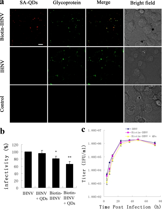
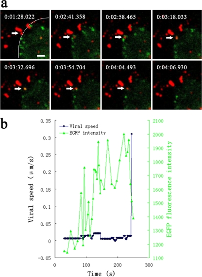
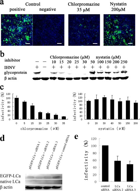
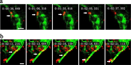
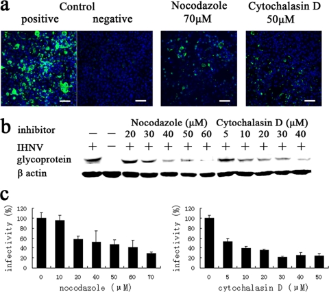
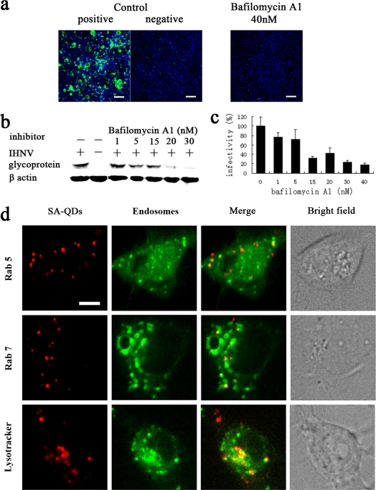
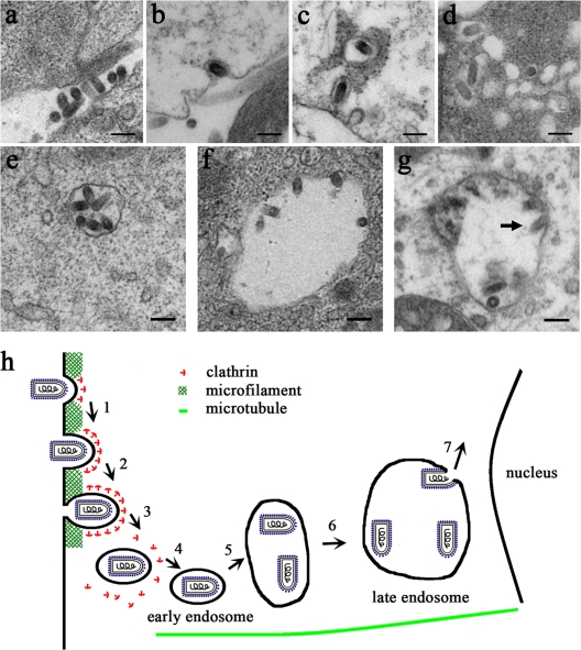
Infectious hematopoietic necrosis virus (IHNV) is an important fish pathogen that infects both wild and cultured salmonids. As a species of the genus Novirhabdovirus, IHNV is a valuable model system for exploring the host entry mechanisms of rhabdoviruses. In this study, quantum dots (QDs) were used as fluorescent labels for sensitive, long-term tracking of IHNV entry. Using live-cell fluorescence microscopy, we found that IHNV is internalized through clathrin-coated pits after the virus binds to host cell membranes. Pretreatment of host cells with chlorpromazine, a drug that blocks clathrin-mediated endocytosis, and clathrin light chain (LCa) depletion using RNA interference both resulted in a marked reduction in viral entry. We also visualized transport of the virus via the cytoskeleton (i.e., actin filaments and microtubules) in real time. Actin polymerization is involved in the transport of endocytic vesicles into the cytosol, whereas microtubules are required for the trafficking of clathrin-coated vesicles to early endosomes, late endosomes, and lysosomes. Disrupting the host cell cytoskeleton with cytochalasin D or nocodazole significantly impaired IHNV infectivity. Furthermore, infection was significantly affected by pretreating the host cells with bafilomycin A1, a compound that inhibits the acidification of endosomes and lysosomes. Strong colocalizations of IHNV with endosomes indicated that the virus is internalized into these membrane-bound compartments. This is the first report in which QD labeling is used to visualize the dynamic interactions between viruses and endocytic structures; the results presented demonstrate that IHNV enters host cells via clathrin-mediated endocytic, cytoskeleton-dependent, and low-pH-dependent pathways.


Figures

Fig. 1.
Fig. 1.
Labeling of IHNVs with QDs. EPC cells were incubated with biotin-IHNV, IHNV, or no virus (control) for 20 min at 4°C. (a) Immunofluorescence staining was performed using antiserum (green) and IHNV particles that were also incubated with SA-QDs (red). Scale bar, 10 μm. (b) The same quantities of biotin-IHNV and IHNV were used at each group, and then cells were either incubated with SA-QDs or left untreated. Viral infectivity was calculated by qPCR analysis at 24 h p.i. Asterisks denote a statistically significant change in infectivity compared to native IHNV (*, P < 0.05; **, P < 0.01). The data shown represent the mean values and standard deviations of the results. Three independent experiments, repeated three times for each sample, were performed. (c) One-step growth curve of IHNV, biotin-IHNV, and QD-labeled IHNV in EPC cells. Initial infections were done at an MOI of 5. Titers in the supernatants at each time of collection were determined in EPC cells.
Fig. 2.
Fig. 2.
Internalization of IHNV particles through clathrin-mediated endocytosis. (a) Snapshots of an IHNV particle (indicated by an arrow) being internalized through de novo formation of a CCP. A movie depicting this process is also available (see Video S1 in the supplemental material). EPC cells were transfected with pEGFP-LCa prior to incubation with QD-labeled IHNVs. The border of the cell is indicated by a curve. The time at which each snapshot was taken is indicated in white at the top of each frame. Scale bar, 2 μm. (b) The time trajectories of an IHNV particle internalized through de novo formation of a CCP. Black symbols represent the velocity-time trajectories of the virus. Green symbols represent the integrated fluorescence intensity of EGFP-LCa associated with the particle. The EGFP fluorescence intensity gradually increased and then rapidly disappeared (corresponding to disassembly of the clathrin coat), followed by rapid movement of the particle.
Fig. 3.
Fig. 3.
Effect of endocytosis inhibitors on infection of EPC cells by IHNV. (a) Cells, either untreated (positive control) or pretreated with either 35 μM chlorpromazine or 200 μM nystatin, were infected with IHNV. Uninfected cells (negative control) were also included in the experiment. At 48 h p.i., immunofluorescence staining was performed using antiserum (green) and Hoechst nuclear stain (blue). Scale bar, 50 μm. (b and c) Cells were treated with various concentrations of chlorpromazine and nystatin prior to infection with IHNV. Viral infection was quantified by Western blot analysis at 48 h p.i. (b) and by qPCR analysis at 24 h p.i. (c). Viral infectivity was quantified as the percentage of amplified viral RNA of drug-treated cells relative to untreated control cells. (d) Cells were cotransfected with siRNAs and pEGFP-LCa, and expression of LCa was detected by Western blot analysis after 48 h. (e) Cells were infected by IHNV after 48 h of transfection of siRNA, and viral infectivity was calculated by qPCR analysis at 24 h p.i. The data shown represent the mean values and standard deviations of the results. At least two independent experiments, repeated three times for each sample, were performed.
Fig. 4.
Fig. 4.
Host cytoskeleton-dependent IHNV entry pathway. Snapshots of IHNV particle (indicated by an arrow) transport via the cytoskeleton (actin filament in panel a and microtubule in panel b). Movies depicting these processes are available (see Videos S2 and S3 in the supplemental material). EPC cells were transfected with pcDNA3.1-GFP-mTn and pcDNA3.1-GFP-MAP4 prior to incubation with QD-labeled IHNVs. The time at which each snapshot was taken is indicated in white at the top of each frame. Scale bar, 2 μm.
Fig. 5.
Fig. 5.
The effects of cytoskeleton-disrupting drugs on the infection of EPC cells by IHNV. (a) Cells, either untreated (positive control) or treated with 70 μM nocodazole or 50 μM cytochalasin D, were infected with IHNV. Uninfected cells (negative control) were also included in the experiment. At 48 h p.i., immunofluorescence staining was performed as described in the legend of Fig. 3a. Scale bar, 50 μm. (b and c) Cells were treated with various concentrations of nocodazole and cytochalasin D prior to infection with IHNV. Viral infectivity was quantified as described in the legends of Fig. 3b and c.
Fig. 6.
Fig. 6.
Low-pH-dependent entry pathway. (a) Cells, either untreated (positive control) or treated with 40 nM bafilomycin A1, were infected with IHNV. Uninfected cells (negative control) were also included in the experiment. At 48 h p.i., immunofluorescence staining was performed as described in the legend of Fig. 3a. Scale bar, 50 μm. (b and c) Cells were treated with various concentrations of bafilomycin A1 prior to infection with IHNV. Viral infectivity was quantified as described in the legends of Fig. 3b and c. (d) EPC cells were transfected with pEGFP-Rab5 and pEGFP-Rab7 prior to incubation with QD-labeled IHNVs. Colocalizations of IHNV with Rab5 and Rab7 were imaged at 5 and 30 min p.i., respectively. Fusion of IHNV particles with early endosomes is also shown in Video S4 in the supplemental material. Cells were inoculated with QD-labeled IHNV. At 30 min p.i. cells were treated with LysoTracker for 5 min. Scale bar, 5 μm.
Fig. 7.
Fig. 7.
Ultrastructural analysis of the entry of IHNVs into EPC cells using electron microscopy. (a) Binding of IHNV particles on the plasma membrane of EPC cells. (b) Uptake of an IHNV particle by a CCP. (c) Internalization of single IHNV particles within CCVs. (d) CCVs containing IHNV particles appear to fuse. (e and f) Large vesicles containing several virus particles were observed. (g) Loosening of the IHNV envelope and fusion with the membrane of an endocytic vesicle membrane (indicated by an arrow). Scale bar, 200 nm. (h) A model of the entry pathway of IHNV into EPC cells. Steps 1 to 3 represent actin filament-dependent active transport: attachment of IHNV to the surfaces of EPC cells and the early stages of clathrin assembly (1), internalization of the viruses via CCPs (2), and the rapid uncoating of CCVs (3). Steps 4 to 6 represent microtubule-dependent movement: fusion with early endosomes (4), fusion of early endosomes with each other (5), and migration into late endosomes and lysosomes (6). Step 7 represents the fusion of IHNV particles with the membranes of endocytotic vesicles and release of its genome into the cytosol for replication.

Similar articles

Cited by

References

    1. Ammayappan A., LaPatra S. E., Vakharia V. N. 2010. Molecular characterization of the virulent infectious hematopoietic necrosis virus (IHNV) strain 220-90. Virol. J. 7:10. - PMC - PubMed
    1. Anderson H. A., Chen Y., Norkin L. C. 1996. Bound simian virus 40 translocates to caveolin-enriched membrane domains, and its entry is inhibited by drugs that selectively disrupt caveolae. Mol. Biol. Cell 7:1825–1834 - PMC - PubMed
    1. Apodaca G. 2001. Endocytic traffic in polarized epithelial cells: role of the actin and microtubule cytoskeleton. Traffic 2:149–159 - PubMed
    1. Bearzotti M., et al. 1999. Fish rhabdovirus cell entry is mediated by fibronectin. J. Virol. 73:7703–7709 - PMC - PubMed
    1. Bucci C., et al. 1992. The small GTPase rab5 functions as a regulatory factor in the early endocytic pathway. Cell 70:715–728 - PubMed

Publication types

MeSH terms