. 2011 Jul;106(1):488-96.
doi: 10.1152/jn.00251.2011. Epub 2011 Apr 27.

Optogenetic photochemical control of designer K+ channels in mammalian neurons

Affiliations

Optogenetic photochemical control of designer K+ channels in mammalian neurons

Doris L Fortin et al. J Neurophysiol. 2011 Jul.

Abstract

Currently available optogenetic tools, including microbial light-activated ion channels and transporters, are transforming systems neuroscience by enabling precise remote control of neuronal firing, but they tell us little about the role of indigenous ion channels in controlling neuronal function. Here, we employ a chemical-genetic strategy to engineer light sensitivity into several mammalian K(+) channels that have different gating and modulation properties. These channels provide the means for photoregulating diverse electrophysiological functions. Photosensitivity is conferred on a channel by a tethered ligand photoswitch that contains a cysteine-reactive maleimide (M), a photoisomerizable azobenzene (A), and a quaternary ammonium (Q), a K(+) channel pore blocker. Using mutagenesis, we identify the optimal extracellular cysteine attachment site where MAQ conjugation results in pore blockade when the azobenzene moiety is in the trans but not cis configuration. With this strategy, we have conferred photosensitivity on channels containing Kv1.3 subunits (which control axonal action potential repolarization), Kv3.1 subunits (which contribute to rapid-firing properties of brain neurons), Kv7.2 subunits (which underlie "M-current"), and SK2 subunits (which are Ca(2+)-activated K(+) channels that contribute to synaptic responses). These light-regulated channels may be overexpressed in genetically targeted neurons or substituted for native channels with gene knockin technology to enable precise optopharmacological manipulation of channel function.


Figures

Fig. 1.
Fig. 1.
Optopharmacological strategy to control the activity of K+ channels. A: the synthetic photoswitch MAQ consists of a cysteine-reactive maleimide, a photoisomerizable azobenzene linker, and a pore-blocking quaternary ammonium (QA; blue). MAQ undergoes trans-to-cis isomerization on illumination with 380-nm light. Exposure to 500-nm light, or prolonged time in darkness, returns the molecule to the trans configuration. B: MAQ covalently attaches to a genetically engineered cysteine located at the appropriate distance from the pore of a K+ channel. C: sequence alignment of different K+ channels. We engineered cysteines at position equivalent to E422C in Kv1.3 (P374C), Kv3.1 (E380C), Kv7.2 (KCNQ; E257C), and small-conductance Ca2+-activated K+ channel type 2 (SK2; Q339C). S5 and S6 denote the 5th and 6th transmembrane domains. P denotes the K+ selectivity filter.
Fig. 2.
Fig. 2.
Engineering a photosensitive Kv1.3. A: introduction of P374C and H401Y and subsequent treatment with MAQ photosensitizes Kv1.3. Whole cell recording from a MAQ-treated HEK-293T cell expressing Kv1.3 P374C and H401Y. The cell was held at −60 mV and stepped to +20 mV every 5 s. The resulting current was measured and plotted over time and wavelength changes. Illumination with 500-nm light blocks the channel, reducing ion flow, whereas illumination with 380-nm light unblocks the channel. B: when the genetically engineered cysteine is protected by treatment with cysteine-reactive reagent MTSET before treatment with MAQ, there is no photosensitization and no regulation of current with light. Kv1.3 channels exhibit cumulative inactivation with very slow recovery (Kupper et al. 2002) that may account for the small decline in currents observed in A and B. C: fraction of current regulated with light in cells expressing Kv1.3 P374C and H401Y treated with MAQ (48.2 ± 16.2%) and pretreated with MTSET to protect the cysteine and prevent attachment of MAQ (0.05 ± 5.5%). The fraction of current photoregulated is defined as the difference between the current under 380- and 500-nm light divided by the amount of current in 380 nm. Data represent average ± SD (n = 6–13).
Fig. 3.
Fig. 3.
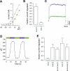
Photosensitization of Kv7.2 channels. A: engineering of a cysteine in Kv7.2 (E257C) and subsequent treatment with MAQ enables photoregulation of the channel. Whole cell recording from a cell expressing Kv7.2 E257C treated with MAQ. Current was elicited by stepping to −40 mV from a holding voltage of −70 mV. Illumination with 500-nm light blocks the channel, whereas the 380-nm light unblocks the channel. B: tail current through Kv7.2 E257C is also photoregulated after treatment with MAQ. Voltage protocol to elicit tail current is the same as in A. C: fraction of current regulated in cells expressing Kv7.2 K255C (−1.4 ± 6.2%), G256C (−0.7 ± 1.1%), E257C (33.8 ± 8.3%), and G259C (−14.2 ± 18.3%). There was no detectable Kv7.2 current in cells expressing the mutant N258C (N/A). Currents were elicited as in A. The position of the engineered cysteine is crucial for photosensitization. Data represent averages ± SD (n = 3–9).
Fig. 4.
Fig. 4.
Photosensitization of Kv3 family channels. A: steady-state current voltage curves from a voltage-clamped HEK-293 cell expressing Kv3.1 E380C. Currents were elicited by stepping to the indicated voltage from a holding potential of −70 mV and measured in 380-nm (open squares; violet) or 500-nm (closed squares; green) light after treatment with MAQ. B: fraction of current photoswitched after MAQ treatment of cells expressing Kv3.1 E380C (67.0 ± 6.5%) or wild-type Kv3.1 (9.2 ± 3.5%). Currents were elicited by stepping from a resting potential of −70 to −10 mV. Data represent average ± SD (n = 3–9). C: representative currents elicited by stepping from a holding potential of −70 to −10 mV in alternating 500- (green), 380- (violet), and 500-nm (green) light. Currents through photosensitive Kv3.1 are repetitively blocked by 500-nm light with little decrement in the fraction of current regulated and no apparent photobleaching of the photoswitch. D: currents were elicited and measured every 2 s as in C and plotted over time and wavelength changes. Illumination with 500-nm light (green) blocks the channel, whereas illumination with 380-nm light (violet) unblocks the channel. E: channels expressed in HeLa cells composed of up to 2 (heteromers) or 4 cysteine (homomers)-containing subunits are photosensitized to a similar extent by MAQ treatment (homomeric Kv3.4, 5.540 ± 3.6%; homomeric Kv3.4 D420C, 57.2 ± 15.9%; heteromeric Kv3.4 D420C + Kv3.1, 49.8 ± 6.0%; heteromeric Kv3.1 E380C, 75.6 ± 24.2%; heteromeric Kv3.1 E380C + Kv3.4, 59.1 ± 12.6%. Data represent average ± SD, n = 4–5 for each combination).
Fig. 5.
Fig. 5.
Photosensitization of Kv3.1 channels in neurons enable control of repetitive firing. A: photoregulation enables control of repetitive firing in a neuron expressing Kv3.1 E380C after MAQ treatment. The number of action potentials fired in response to sustained current injection (iinj) is higher under 380-nm (violet) than 500-nm (green) light. Light exposure did not affect resting membrane potential. B: photoregulation of Kv3.1 E380C enables control of action potential firing in transfected neurons. Six of seven cells expressing Kv3.1 E380C and treated with MAQ fire more action potentials in 380-nm light in response to a depolarizing current injection. C: photoregulation of Kv3.1 E380C changes the shape of the action potential in transfected neurons treated with MAQ. Single action potentials were generated by short (10 ms) current injection. The afterhyperpolarization is more pronounced under 380-nm (violet) than in 500-nm (green) light.
Fig. 6.
Fig. 6.
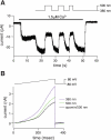
Photosensitization of SK2 channels. A: inside-out patch recording from a HEK-293T cell expressing SK2 Q339C and treated with MAQ. Ca2+-activated currents were induced with a solution containing 1.5 μM free Ca2+ buffered with EGTA. Ca2+-activated current is larger in 380- than 500-nm light. B: whole cell recording showing light-sensitive currents during a depolarizing voltage ramp (−80 to +80 mV). The pipette contained 1 μM Ca2+ to ensure maintained activation of SK2 channels. Currents were maximal in 380-nm light and reduced in 500-nm light. Apamin (0.5 μM) further inhibited the current, indicating that blockade by trans MAQ was >60% complete.
Fig. 7.
Fig. 7.
Photoregulation of SK2 channels in hippocampal slices. A: whole cell recordings from hippocampal CA1 neurons expressing SK2 Q339C in a MAQ-treated slice. A depolarizing pulse (top) elicited tail currents (bottom) that were larger in 380-nm (green) than in 500-nm light (violet). B: light has no effect on currents measured from a nonexpressing cell in a MAQ-treated slice. C: summary data for photocontrol of the SK-mediated tail current. On average, 500-nm light reduced the tail current measured 100 ms after the pulse by 30 ± 19% (n = 12; average ± SD) for cells expressing SK2 Q339C and by 3 ± 9% (n = 14; average ± SD) for nonexpressing cells. *Significant difference (P < 0.01; paired t-test). D: average excitatory postsynaptic potentials (EPSP) waveform from a SK2 Q339C-expressing neuron in 500-nm light (green), which blocks the SK2 channels, and 380-nm light (violet), which unblocks the channels. E: repeated changes in peak EPSP amplitude with 500- and 380-nm light.

References

    1. Abbott GW. Molecular mechanisms of cardiac voltage-gated potassium channelopathies. Curr Pharm Des 12: 3631–3644, 2006 - PubMed
    1. Banghart M, Borges K, Isacoff E, Trauner D, Kramer RH. Light-activated ion channels for remote control of neuronal firing. Nat Neurosci 7: 1381–1386, 2004 - PMC - PubMed
    1. Baranauskas G, Tkatch T, Nagata K, Yeh JZ, Surmeier DJ. Kv3.4 subunits enhance the repolarizing efficiency of Kv3.1 channels in fast-spiking neurons. Nat Neurosci 6: 258–266, 2003 - PubMed
    1. Beeton C, Wulff H, Standifer NE, Azam P, Mullen KM, Pennington MW, Kolski-Andreaco A, Wei E, Grino A, Counts DR, Wang PH, LeeHealey CJ, Andrews B S, Sankaranarayanan A, Homerick D, Roeck WW, Tehranzadeh J, Stanhope KL, Zimin P, Havel PJ, Griffey S, Knaus HG, Nepom GT, Gutman GA, Calabresi PA, Chandy KG. Kv1.3 channels are a therapeutic target for T cell-mediated autoimmune diseases. Proc Natl Acad Sci USA 103: 17414–17419, 2006 - PMC - PubMed
    1. Blaustein RO, Cole PA, Williams C, Miller C. Tethered blockers as molecular ‘tape measures’ for a voltage-gated K+ channel. Nat Struct Biol 7: 309–311, 2000 - PubMed

Publication types

MeSH terms

LinkOut - more resources