. 2011 Jun 1;186(11):6585-96.
doi: 10.4049/jimmunol.1002794. Epub 2011 Apr 27.

Staphylococcus aureus biofilms prevent macrophage phagocytosis and attenuate inflammation in vivo

Affiliations

Staphylococcus aureus biofilms prevent macrophage phagocytosis and attenuate inflammation in vivo

Lance R Thurlow et al. J Immunol. .

Abstract

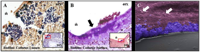
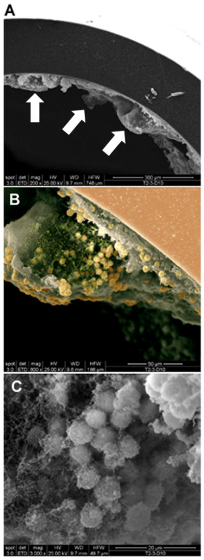
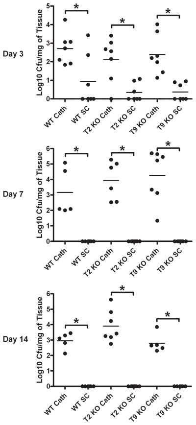
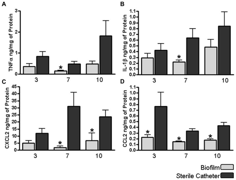
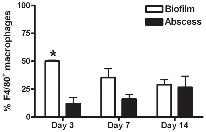
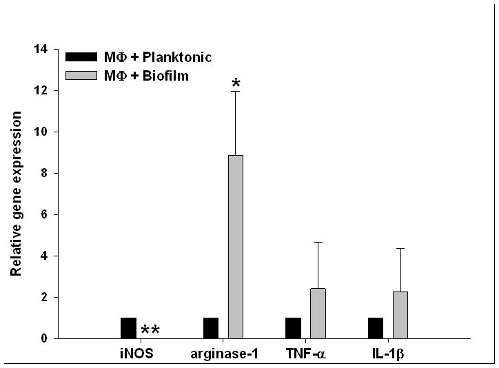
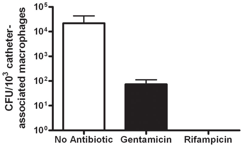
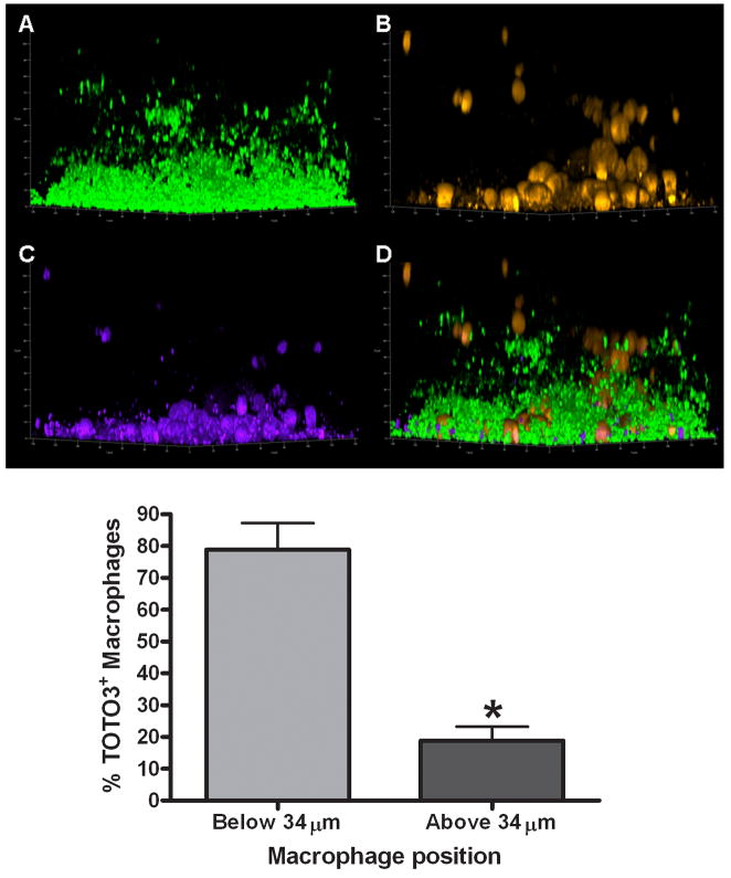
Biofilms are complex communities of bacteria encased in a matrix composed primarily of polysaccharides, extracellular DNA, and protein. Staphylococcus aureus can form biofilm infections, which are often debilitating due to their chronicity and recalcitrance to antibiotic therapy. Currently, the immune mechanisms elicited during biofilm growth and their impact on bacterial clearance remain to be defined. We used a mouse model of catheter-associated biofilm infection to assess the functional importance of TLR2 and TLR9 in the host immune response during biofilm formation, because ligands for both receptors are present within the biofilm. Interestingly, neither TLR2 nor TLR9 impacted bacterial density or inflammatory mediator secretion during biofilm growth in vivo, suggesting that S. aureus biofilms circumvent these traditional bacterial recognition pathways. Several potential mechanisms were identified to account for biofilm evasion of innate immunity, including significant reductions in IL-1β, TNF-α, CXCL2, and CCL2 expression during biofilm infection compared with the wound healing response elicited by sterile catheters, limited macrophage invasion into biofilms in vivo, and a skewing of the immune response away from a microbicidal phenotype as evidenced by decreases in inducible NO synthase expression concomitant with robust arginase-1 induction. Coculture studies of macrophages with S. aureus biofilms in vitro revealed that macrophages successful at biofilm invasion displayed limited phagocytosis and gene expression patterns reminiscent of alternatively activated M2 macrophages. Collectively, these findings demonstrate that S. aureus biofilms are capable of attenuating traditional host proinflammatory responses, which may explain why biofilm infections persist in an immunocompetent host.


Figures

Figure 1
Figure 1. Characterization of S. aureus localization during biofilm development
Catheters and surrounding tissues were isolated from wild type mice at day 10 following S. aureus infection and subjected to Gram-staining (A and B) or SEM (C). There was evidence of bacterial growth (purple) within the catheter lumen (A) as well as host tissue immediately surrounding the catheter (B, arrow). Insets in panels A & B depict 10X magnification with red rectangles indicating the area enlarged at 40X magnification and asterisks denoting the original location of infected catheters that were non-adherent to glass slides. A 400X magnification SEM image (C) has been pseudocolored to highlight S. aureus biofilm (blue) at the interface between the catheter (grey) and surrounding host tissue (purple). Host cells located at a distance from the biofilm are indicated by arrows.
Figure 2
Figure 2. Evidence of catheter-associated biofilm growth in vivo
Catheters were isolated from wild type mice at day 10 following S. aureus infection and processed for SEM analysis. The smooth surface at the periphery of the images in A and B represents the catheter with biofilm visible on the internal face. (A), 200X magnification demonstrating the irregular undulating pattern of the biofilm surface (arrows indicate tower structures); (B), higher magnification of a tower from the image shown in (A) revealing a predominantly hollow interior with numerous cocci at the margins (800X magnification). The image has been pseudocolored to highlight S. aureus (gold) and presumably matrix material (green), which likely aggregated during the SEM dehydration process, from the remaining biofilm structure (grey; 800X magnification). The smooth surface at the upper right represents the catheter (salmon) with biofilm visible on the internal face. (C), 3000X magnification of a S. aureus cluster within the tower depicted in (B).
Figure 3
Figure 3. Visualization of S. aureus biofilm infection using IVIS
Wild type (WT), TLR2 knockout (KO), and TLR9 KO mice were infected with 5 × 10⁵ CFU of USA300 LAC::lux either in the lumen of surgically implanted catheters to establish biofilm infection or subcutaneously in the absence of any indwelling device. At the indicated time points post-infection, mice were subjected to IVIS imaging to visualize the extent of biofilm or subcutaneous infection. Images are presented from one mouse per group that was imaged at all time points to map the progression of biofilm infection.
Figure 4
Figure 4. Infection with an equivalent S. aureus dose leads to the establishment of catheter-related biofilm infection but rapid clearance from subcutaneous sites
Wild type (WT), TLR2 knockout (KO), and TLR9 KO mice were infected with 5 × 10⁵ CFU of USA300 LAC::lux either in the lumen of surgically implanted catheters (cath) to establish biofilm infection or subcutaneously (sc) in the absence of any indwelling device. Animals were sacrificed at the indicated days following S. aureus infection, whereupon host tissues surrounding infected catheters or s.c. injection sites were homogenized to quantitate bacterial burdens. Results are expressed as the number of CFU per mg host tissue to correct for differences in tissue sampling size. Significant differences in bacterial burdens between biofilm-infected versus s.c. injected mice are denoted by asterisks (*, p < 0.001).
Figure 5
Figure 5. S. aureus biofilm attenuates inflammatory mediator expression compared to the wound healing response elicited following sterile catheter implantation
Tissues surrounding S. aureus infected or sterile catheters from wild type mice were collected and homogenized at the indicated time points to obtain supernatants to quantitate differences in TNF-α (A), IL-1β (B), CXCL2 (C), or CCL2 (D) expression by ELISA. Results were normalized to the amount of total protein recovered to correct for differences in tissue sampling size. Significant differences in inflammatory mediator expression between biofilm infected versus sterile catheters are denoted by asterisks (*, p < 0.05). Results are presented from individual animals combined from three independent experiments.
Figure 6
Figure 6. S. aureus biofilms elicit exaggerated macrophage infiltrates compared to abscesses
Wild type mice were infected with 5 × 10⁵ CFU of USA300 LAC::lux either in the lumen of surgically implanted catheters or subcutaneously in the absence of any indwelling device to establish biofilm and abscess infections, respectively. Animals were sacrificed at days 3, 7, or 14 following S. aureus exposure, whereupon tissues surrounding infected catheters or s.c. injection sites were collected to quantitate macrophage infiltrates by FACS. Results are expressed as the percent of F4/80+ macrophages after correction for isotype control staining and represent the mean ± SEM of three independent experiments. Significant differences in macrophage infiltration into biofilm versus abscess infections are denoted by asterisks (*, p < 0.01).
Figure 7
Figure 7. Macrophage migration towards S. aureus biofilms in vivo is limited
Tissues surrounding biofilm-infected catheters were collected from wild type mice at day 7 following S. aureus infection and subjected to immunofluorescence staining with Iba-1 to identify macrophages (red) and DAPI to demarcate nuclei (blue) by confocal microscopy (20X magnification). Asterisks indicate the location of infected catheters and the border between biofilm and surrounding host tissue is denoted by dashed lines. Results are representative of 6 individual animals.
Figure 8
Figure 8. S. aureus biofilms repress iNOS and augment arginase-1 expression
Wild type mice were infected with 5 × 10⁵ CFU of USA300 LAC::lux either in the lumen of surgically implanted catheters or subcutaneously in the absence of any indwelling device to establish biofilm and abscess infections, respectively. Animals were sacrificed at day 7 following S. aureus exposure, whereupon tissues surrounding infected catheters or s.c. injection sites were collected and subjected to immunofluorescence staining with arginase-1 (red) and DAPI to identify nuclei (blue) by confocal microscopy (20X magnification). Stars represent the location of material remaining from the catheter lumen and the diamonds denote the necrotic abscess core. The border between biofilm and surrounding host tissue is depicted by arrows. Results are representative of 6 individual animals.
Figure 9
Figure 9. A subset of biofilm-associated macrophages express arginase-1
Tissues surrounding biofilm-infected catheters were collected from wild type mice at day 7 following S. aureus infection and subjected to immunofluorescence staining for the macrophage-specific marker Iba-1 (red) and arginase-1 (green) to determine co-localization patterns by confocal microscopy (arrows, 20X magnification). The position of the biofilm-infected catheter is denoted by asterisks. Results are representative of 6 individual animals.
Figure 10
Figure 10. S. aureus biofilms skew macrophage gene expression towards an alternatively activated (M2) phenotype
Bone marrow-derived macrophages from GFP Tg mice were incubated with 6 day-old biofilms or planktonic bacteria for 2 h, whereupon viable macrophages were purified by FACS and RNA immediately isolated for qRT-PCR analysis. Gene expression levels in macrophages exposed to S. aureus biofilms were calculated after normalizing signals against GAPDH and are presented as the fold-change relative to macrophages incubated with planktonic bacteria. Results represent the mean ± SEM of four independent experiments (*, p < 0.05; **, p < 0.001).
Figure 11
Figure 11. Macrophages actively phagocytize planktonic S. aureus but not biofilm-associated bacteria
(A) Primary macrophages labeled with CellTracker orange (orange) were exposed to 4 day-old USA300 LAC-GFP biofilms or planktonic bacteria (green) for a 24 h period, whereupon visualization of intracellular bacteria was evaluated by confocal microscopy (63X magnification, 1 μm slice). Biofilm-associated macrophages exhibited little evidence of phagocytosis, whereas intracellular bacteria were readily detectable in macrophages incubated with planktonic organisms. Results are representative of six independent experiments. In (B), four and six day-old USA300 LAC-GFP static biofilms were mechanically disrupted by triturating and incubated with macrophages for 1 h. Planktonic bacteria were included as a positive control. Fluorescent microscopy of cytospin preparations shows the ability of macrophages to phagocytize planktonic (left) as well as disassociated bacteria from four day-(center) and six day-old (right) biofilms. Results are representative of three independent experiments.
Figure 12
Figure 12. Macrophages recovered from S. aureus biofilm infections harbor viable intracellular bacteria
Viable macrophages (F4/80+, 7-AAD) were recovered from tissues surrounding biofilm infections at day 7 post-infection by FACS, whereupon gentamicin protection assays were performed to evaluate the presence of viable intracellular bacteria. Controls included sorted macrophages without any antibiotic treatment (to assess extra- and intracellular bacteria) and macrophages treated with rifampicin, which is capable of penetrating the eukaryotic cell membrane. Results are expressed as the number of viable S. aureus (CFU per 103 biofilm-associated macrophages) and represent the mean ± SEM of five independent experiments.
Figure 13
Figure 13. Macrophages exposed to mature S. aureus biofilm exhibit a dystrophic morphology
Primary macrophages labeled with CellTracker orange (CTO; yellow-orange) were exposed to 4 or 6 day-old USA300 LAC-GFP biofilms (green) for a 24 h period, whereupon visualization of macrophage morphology was evaluated by confocal microscopy (63X magnification, 1 μm slice). Macrophages infiltrating immature (4 day-old) biofilms retained a typical rounded morphology, whereas macrophages infiltrating mature biofilms (6 day-old) often exhibited a dystrophic ghost-like morphology with minimal retention of CTO (arrows). Results are representative of six independent experiments.
Figure 14
Figure 14. Macrophage viability is dictated by invasion into the biofilm proper
(A) 4 day-old USA300 LAC-GFP biofilms (average height ~ 33 μm) were incubated with (B) primary macrophages labeled with CellTracker orange (yellow-orange) for a 24 h period, whereupon macrophage viability was determined by uptake of the live/dead stain TOTO3 (purple; C). The majority of dead (TOTO3+) macrophages are within 34 μm of the biofilm proper (p < 0.05) and a composite image of all staining is shown (D). Bottom; significant differences between the percentages of TOTO3+ macrophages based on location within the biofilm proper are denoted with an asterisk (*, p < 0.05; n = 218 cells).
Figure 15
Figure 15. S. aureus biofilms skew macrophage responses to favor bacterial persistence
Despite the presence of TLR2 and TLR9 ligands within the biofilm (lipoproteins/PGN and eDNA, respectively), these traditional pattern recognition receptors to not impart macrophage responsiveness or bacterial clearance during biofilm infection (depicted by red “x” marks). Likewise, the expression of iNOS is reduced following biofilm formation concomitant with an increase in arginase-1. Collectively, these changes shift the immune response away from a bactericidal pathway and likely contribute to biofilm persistence.

References

    1. Donlan RM, Costerton JW. Biofilms: survival mechanisms of clinically relevant microorganisms. Clin Microbiol Rev. 2002;15:167–193. - PMC - PubMed
    1. Otto M. Staphylococcal biofilms. Curr Top Microbiol Immunol. 2008;322:207–228. - PMC - PubMed
    1. Fitzpatrick F, Humphreys H, O’Gara JP. The genetics of staphylococcal biofilm formation--will a greater understanding of pathogenesis lead to better management of device-related infection? Clin Microbiol Infect. 2005;11:967–973. - PubMed
    1. Burlak C, Hammer CH, Robinson MA, Whitney AR, McGavin MJ, Kreiswirth BN, Deleo FR. Global analysis of community-associated methicillin-resistant Staphylococcus aureus exoproteins reveals molecules produced in vitro and during infection. Cell Microbiol. 2007;9:1172–1190. - PMC - PubMed
    1. Chambers HF, Deleo FR. Waves of resistance: Staphylococcus aureus in the antibiotic era. Nat Rev Microbiol. 2009;7:629–641. - PMC - PubMed

Publication types

MeSH terms