. 2011 Jun 1;186(11):6227-37.
doi: 10.4049/jimmunol.1002950. Epub 2011 Apr 27.

The pleckstrin homology domain in the SKAP55 adapter protein defines the ability of the adapter protein ADAP to regulate integrin function and NF-kappaB activation

Affiliations

The pleckstrin homology domain in the SKAP55 adapter protein defines the ability of the adapter protein ADAP to regulate integrin function and NF-kappaB activation

Brandon J Burbach et al. J Immunol. .

Abstract

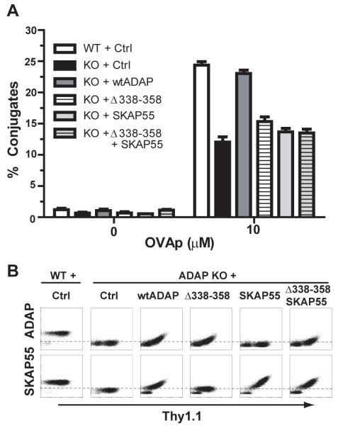
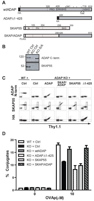
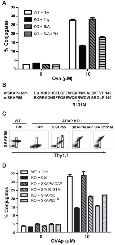
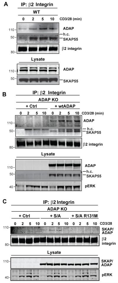
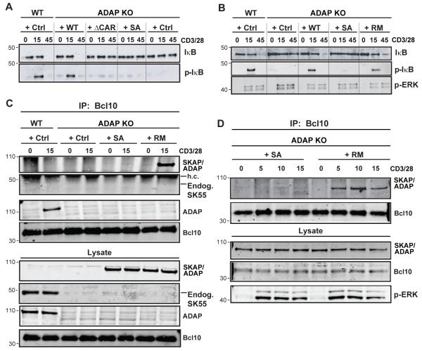
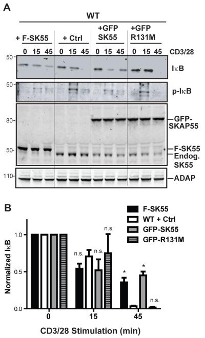
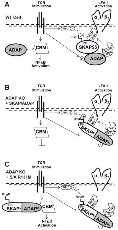
Adhesion and degranulation promoting adapter protein (ADAP) is a multifunctional hematopoietic adapter protein that regulates TCR-dependent increases in both integrin function and activation of the NF-κB transcription factor. Activation of integrin function requires both ADAP and the ADAP-associated adapter Src kinase-associated phosphoprotein of 55 kDa (SKAP55). In contrast, ADAP-mediated regulation of NF-κB involves distinct binding sites in ADAP that promote the inducible association of ADAP, but not SKAP55, with the CARMA1 adapter and the TAK1 kinase. This suggests that the presence or absence of associated SKAP55 defines functionally distinct pools of ADAP. To test this hypothesis, we developed a novel SKAP-ADAP chimeric fusion protein and demonstrated that physical association of ADAP with SKAP55 is both sufficient and necessary for the rescue of integrin function in ADAP-deficient T cells. Similar to wild-type ADAP, the SKAP-ADAP chimera associated with the LFA-1 integrin after TCR stimulation. Although the SKAP-ADAP chimera contains the CARMA1 and TAK1 binding sequences from ADAP, expression of the chimera does not restore NF-κB signaling in ADAP(-/-) T cells. A single point mutation in the pleckstrin homology domain of SKAP55 (R131M) blocks the ability of the SKAP-ADAP chimera to restore integrin function and to associate with LFA-1. However, the R131M mutant was now able to restore NF-κB signaling in ADAP-deficient T cells. We conclude that integrin regulation by ADAP involves the recruitment of ADAP to LFA-1 integrin complexes by the pleckstrin homology domain of SKAP55, and this recruitment restricts the ability of ADAP to interact with the NF-κB signalosome and regulate NF-κB activation.


Figures

Figure 1
Figure 1. Impaired conjugate efficiency in the absence of ADAP and SKAP55 interaction
(A) Naïve hCAR/DO11.10 (WT) or hCAR/DO11.10/ADAP−/− (KO) CD4 T cells were transduced with control (Ctrl) Thy1.1 adenovirus or the indicated ADAP or SKAP55 expression construct(s). After 3d, the transduced cells were incubated with OVAp-pulsed splenic B cells and analyzed by flow cytometry for efficiency of T:APC conjugate formation as described in Material and Methods. (B) Intracellular staining for ADAP or SKAP55 was performed on the cells transduced in (A), and plotted versus Thy1.1 expression. Conjugates depicted in A were gated on the top one-third of the Thy1.1 gate to ensure that SKAP55 and/or ADAP levels were comparable to endogenous SKAP55 and/or ADAP expression in wild-type control cells. Results for both the conjugate assays and the intracellular staining are representative of at least 3 independent experiments.
Figure 2
Figure 2. A SKAP/ADAP chimeric fusion protein restores T:APC conjugate formation in ADAP−/− T cells
(A) Schematic diagram of key expression constructs used in this study. Amino acid numbering is given for the murine ADAP p130kDa isoform and for human SKAP55. PRO, proline-rich domain; E/K, glutamic acid and lysine-rich domain; hSH3N or hSH3C, N-terminal or C-terminal helical SH3 domain; PH, pleckstrin homology domain. Asterisks indicate the positions of key tyrosine residues (547/549/584/615/687) that have been reported for SLP-76 and Fyn binding to ADAP. The SKAP/ADAP chimera is a 692 aa molecule comprised of the N terminal 299 aa of human SKAP55 fused to the C-terminus of murine ADAP beginning at aa426. (B) Western blot analysis of SKAP55 and ADAP expression in whole cell lysates prepared from naïve wild-type (WT) or ADAP−/− (KO) cells expressing the indicated construct. (C) Naïve hCAR/DO11.10 (WT) or hCAR/DO11.10/ADAP−/− (KO) T cells were transduced for 3d with adenovirus for the indicated construct, fixed, and analyzed by intracellular staining for ADAP (N-terminus), SKAP55, or the HA-Tag. (D) Conjugate assays were performed as in Figure 1. The conjugate efficiency was evaluated from the Thy1.1 gated boxes depicted in panel C, to ensure that the level of the SKAP/ADAP chimera and the control constructs matched that of endogenous SKAP55 observed in wild-type control cells. Results are representative of at least 4 independent experiments for each construct.
Figure 3
Figure 3. The R131 amino acid residue in the SKAP55 PH domain is critical for efficient ADAP-dependent T:APC conjugate formation
(A) Naïve hCAR/DO11.10 (WT) or hCAR/DO11.10/ADAP−/− (KO) T cells were transduced with the indicated constructs and conjugate assays were performed as described in Figures 1-2. The SKAP/ADAPΔPH construct (S/AΔPH) lacks the entire 100 aa PH domain of SKAP55. (B) Amino acid alignment of SKAP-HOM and SKAP55 depicting the lipid binding region of the PH domain. Arginine 140 in SKAP-HOM corresponds to R131 in SKAP55. (C) Expression of SKAP55, SKAP/ADAP, or SKAP/ADAP R131M (S/A R131M) in naïve hCAR/DO11.10/ADAP−/− T cells. Note that the highest levels of SKAP55 expression achieved in the absence of ADAP (Thy1.1hi cells; boxed gate marked “Hi” in middle panel) approach the levels of endogenous SKAP55 found in naïve wild-type T cells, and correspond to the SKAP55 level found in Thy1.1lo cells expressing the SKAP/ADAP chimera or the SKAP/ADAP R131M mutant. (D) Conjugate assays were performed as described in Figures 1-2. T cells were gated on Thy1 lo cells (leftmost gate on each plot in C) to match endogenous levels of SKAP55 found in wild-type cells. For comparison, the conjugate efficiency of Thy1.1hi cells following SKAP55 expression alone in ADAP−/− is also shown (SKAP55Hi). Similar results were obtained in at least 4 independent experiments.
Figure 4
Figure 4. The SKAP55 PH domain is required for TCR-dependent recruitment to β2 integrins
(A) hCAR/DO11.10 (WT) CD4 T cell blasts were stimulated for the indicated times with anti-CD3/CD28. The cells were lysed and subjected to anti-β2 (CD18) integrin immunoprecipitation. Western blots were probed for ADAP, SKAP55 and for the β2 integrin. Whole cell lysate inputs (lower 2 panels) are shown to demonstrate ADAP and SKAP55 expression. (B,C) hCAR/DO11.10/ADAP−/− (ADAP KO) T cell blasts were transduced with the indicated adenovirus constructs and stimulated and immunoprecipitated for β2 integrin as in (A). Western blots were probed with antibodies against ADAP and SKAP55 to detect ADAP and SKAP55 (B) or the SKAP/ADAP chimera (C), and with antibodies against the β2 integrin to detect the immunoprecipitated CD18 integrin subunit. Whole cell lysate inputs (lower panels) are shown to demonstrate expression of ADAP, SKAP55, and the SKAP/ADAP chimera, and activation was confirmed by blotting for phosphoERK. Similar results were observed in 4 independent experiments.
Figure 5
Figure 5. The SKAP55 PH domain inhibits ADAP-dependent NF-κB activation
(A) Naïve hCAR/DO11.10 (WT) and hCAR/DO11.10/ADAP−/− (ADAP KO) T cells were transduced with control Thy1.1 adenovirus or adenovirus expressing wild-type ADAP (WT), ADAPΔCAR (ΔCAR), or SKAP/ADAP chimera (S/A). After 3d, cells were stimulated with anti-CD3/CD28 for the indicated times (minutes) and lysates prepared. Western blots were probed with antibodies to total IκBα and phospho-IκBα to demonstrate IκBα degradation and phosphorylation, respectively. (B) Western blots from hCAR/DO11.10 and hCAR/DO11.10/ADAP−/− T cells expressing the indicated constructs were performed as in A. RM; SKAP/ADAP R131M mutant. (C) T cells were transduced and stimulated for 15 min as in (A-B) and subjected to immunoprecipitation with mouse anti-Bcl10 antibodies. Immune complexes were analyzed by Western blot with rabbit anti-SKAP55 antibody to detect the appearance of the SKAP/ADAP chimera in the Bcl10 complex. Whole cell lysates are shown in the lower 3 panels and were probed with anti-SKAP55 (to detect the SKAP/ADAP chimera), anti-ADAP, or rabbit anti-Bcl10. (D) T cells from hCAR/DO11.10/ADAP−/− mice were transduced with the indicated constructs as in (C) and stimulated for the indicated times followed by Bcl10 immunoprecipitation and western blotting as described for (C). Similar results were obtained in 3 independent experiments for each panel.
Figure 6
Figure 6. The SKAP55 PH domain suppresses endogenous NF-κB activation
(A) Naïve wild-type hCAR T cells were transduced with adenoviruses to overexpress SKAP55 (F-SK55), GFP-SKAP55 (GFP SK55), or GFP-SKAP55-R131M (GFP R131M). Upper 2 panels, Western blots were performed as in A and B to detect total IκBα or phosphorylated IκBα (p-IκB). Lower 2 panels, Western blots for SKAP55 and ADAP are depicted showing expression of endogenous SKAP55 (Endog. SK55), FLAG-SKAP55 (F-SKAP55), or GFP-SKAP55 (GFP-SKAP55) and equal expression of ADAP in all samples. Results are representative of 3 independent assays performed. (B) Quantification of total IκBα degradation following CD3/CD28 stimulation as described in A. Results are normalized to IκBα densitometry values from the unstimulated samples, and averaged between 3 independent experiments performed. Asterisk (*) indicates P < 0.05 compared to the wild-type control condition within each time point using Bonferroni’s multiple comparison test of the one-way ANOVA. n.s., P>0.05.
Figure 7
Figure 7. Model for ADAP and SKAP55 control of LFA-1 integrin activation and NF-κB activation
(A) In wild-type T cells, TCR stimulation promotes ADAP phosphorylation (P) and activation of the constitutively associated ADAP:SKAP55 adapter module. Positively charged arginine 131 (R131) in the SKAP55 pleckstrin homology (PH) domain recruits and restricts the complex to negatively charged phosphoinositides (such as PIP3) in the plasma membrane. Downstream integrin activation components including Rap1 and a putative Rap1-GEF are provided through ADAP and the Rap1 effectors RIAM and/or RapL through SKAP55 are recruited to LFA-1 for promotion of integrin activation. Only free excess ADAP is available for concurrent assembly of the CARMA-1/Bcl10/Malt1 (CBM) complex, which initiates IκB phosphorylation/degradation and release of NFκB to the nucleus. (B) The SKAP/ADAP chimera is exclusively recruited to the integrin pathway. As there is no excess ADAP that is not associated with SKAP55 in ADAP−/− T cells expressing the chimera, no free ADAP is available for NF-κB activation. (C) In ADAP−/− cells expressing the R131M mutation in the SKAP/ADAP chimera, SKAP55 PH domain-mediated interaction of the chimera with membrane lipids is attenuated. This allows the chimera (through the ADAP C-terminus) to engage the CBM complex and promote NF-κB activation.

References

    1. Burbach BJ, Medeiros RB, Mueller KL, Shimizu Y. T cell receptor signaling to integrins. Immunol. Rev. 2007;218:65–81. - PubMed
    1. Dustin ML, Tseng SY, Varma R, Campi G. T cell-dendritic cell immunological synapses. Curr. Opin. Immunol. 2006;18:512–516. - PubMed
    1. Kane LP, Lin J, Weiss A. It’s all Rel-ative: NF-κB and CD28 costimulation of T-cell activation. Trends Immunol. 2002;23:413–420. - PubMed
    1. Vallabhapurapu S, Karin M. Regulation and function of NF-κB transcription factors in the immune system. Annu. Rev. Immunol. 2009;27:693–733. - PubMed
    1. Koretzky GA, Abtahian F, Silverman MA. SLP76 and SLP65: complex regulation of signalling in lymphocytes and beyond. Nat. Rev. Immunol. 2006;6:67–78. - PubMed

Publication types

MeSH terms