. 2011 Jun 1;186(11):6437-44.
doi: 10.4049/jimmunol.1003897. Epub 2011 Apr 27.

Somatically diversified and proliferating transitional B cells: implications for peripheral B cell homeostasis

Affiliations

Somatically diversified and proliferating transitional B cells: implications for peripheral B cell homeostasis

Venkata A Yeramilli et al. J Immunol. .

Abstract

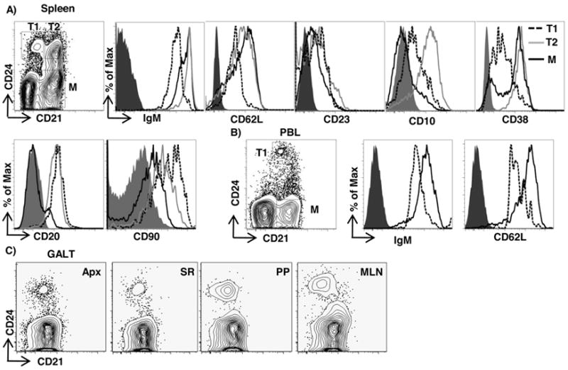
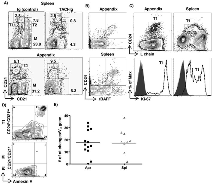
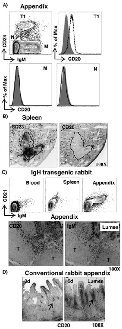
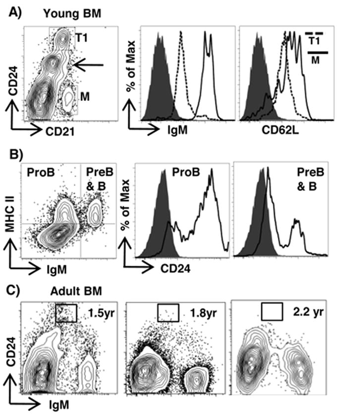
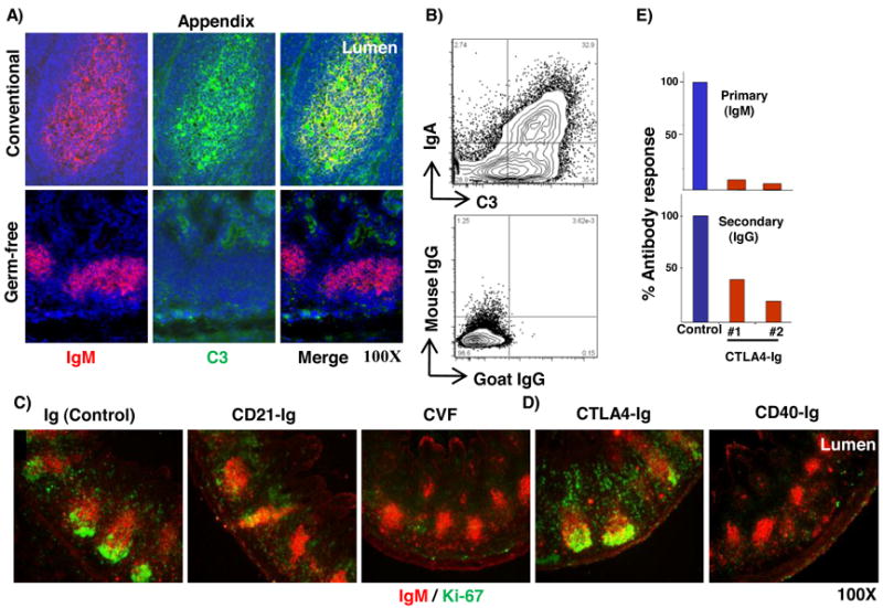
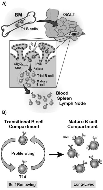
The peripheral B cell compartment in mice and humans is maintained by continuous production of transitional B cells in the bone marrow. In other species, however, including rabbits, B lymphopoiesis in the bone marrow abates early in life, and it is unclear how the peripheral B cell compartment is maintained. We identified transitional B cells in rabbits and classified them into T1 (CD24(high)CD21(low)) and T2 (CD24(high)CD21(+)) B cell subsets. By neutralizing B cell-activating factor in vivo, we found an arrest in peripheral B cell development at the T1 B cell stage. Surprisingly, T1 B cells were present in GALT, blood, and spleen of adult rabbits, long after B lymphopoiesis was arrested. T1 B cells were distinct from their counterparts in other species because they are proliferating and the Ig genes are somatically diversified. We designate these newly described cells as T1d B cells and propose a model in which they develop in GALT, self renew, continuously differentiate into mature B cells, and thereby maintain peripheral B cell homeostasis in adults in the absence of B lymphopoiesis.


Figures

Figure 1
Figure 1. Flow cytometric identification of transitional B cell subsets
A) Staining of splenic B cells for T1 (CD24hiCD21lo) (dashed), T2 (CD24hiCD21+) (gray) and mature (M) (CD24lo/-CD21+) (black) for IgM, CD62L, CD23, CD10, CD38, CD20 and CD90. B) Staining of peripheral blood (PB) T1 (dashed) and M (black) for IgM and CD62L. C) IgM+ B cells from appendix (Apx), sacculus rotundus (SR), Peyer’s patches (PP) and mesenteric lymph nodes (MLN) stained for CD24 and CD21. The gray filled histograms in A and B represent staining with an appropriate isotype control mAb. The plots are representative of staining obtained from at least 3 rabbits.
Figure 2
Figure 2. Functional analysis of transitional B cells
A) Flow cytometric analysis of appendix and spleen cells from TACI-Ig-treated and control (Ig)-treated rabbits stained with anti-CD21 and anti-CD24 mAb. Flow cytometric analysis of appendix and spleen cells from conventional rabbits stained with B) Anti-CD24 and recombinant soluble BAFF (rBAFF) and C) Upper: anti-CD24 and anti-L chain; lower: anti-Ki67 (open histograms) of T1 cells (from upper diagram). Shaded histogram = isotype control. D) Flow cytometric analysis of sorted splenic T1(CD24hiCD21lo) (upper) and mature B cells (CD24lo/-CD21+) (lower) stained with Annexin V and propidium iodide (PI) after 12-15 hrs in culture with anti-Ig (10μg/ml) [goat (F(ab’) anti-rabbit IgG (H+L); Jackson ImmunoResearch Laboratories], E) Somatic diversification of VH regions of PCR-amplified VDJ genes from splenic (spl) and appendix (apx) T1 B cells. The horizontal bar represents average number of nucleotide changes/VH gene (excluding D and J regions); each dot represents one VH gene sequence. Sequences obtained from three adult rabbits are shown. Data in B) to D) are representative of 2-3 independent experiments. Data in A) are representative of two control and three TACI-Ig treated rabbits.
Figure 3
Figure 3. Tissue localization of CD20+ transitional B cells
A) Flow cytometric staining of appendix B cells from a neonatal rabbit (1 wk of age) for T1 (CD24hiIgMlo) (dashed), M (CD24-IgM+) (black) and non-B cells (N) (IgM-CD24-) (black) for CD20. Shaded histogram = isotype control. B) Immunohistological staining of spleen (6-week old) section for CD23 (follicular B cells) and CD20 (transitional B cells). The dotted line represents a B cell follicle. C) Flow cytometric and immunohistological analyses of tissues from an IgH transgenic rabbit stained for IgM and CD21 (upper), and CD20 and IgM (lower), respectively. D) Staining for CD20 in appendix from a conventional 3 & 6-day-old rabbit. Magnification = 100X.
Figure 4
Figure 4. Flow cytometric analysis of T1 B cells in BM
A) BM cells from a young rabbit (8 weeks-of-age) stained for CD21 and CD24, with histograms of IgM and CD62L staining of cells in T1 (dashed) and M (black) cell gates. Shaded histogram = isotype control. B) BM cells from a young rabbit (8 weeks-of-age) stained for IgM, MHC II (left) and CD24, with CD24 histograms of cells in proB and preB & B cell gates (center and right). Shaded histogram = isotype control. C) BM cells from adult rabbits aged 1.5 to 2.2 years stained for CD24 and IgM. All plots are representative of staining obtained from at least 3 rabbits.
Figure 5
Figure 5. Identification of molecules required for proliferative expansion of B cells in GALT
A) Immunofluorescent staining for IgM and C3 in appendix sections from conventional 4 wk old rabbit (upper) and 4 wk old rabbit with a germ-free appendix (lower). B) Flow cytometric analysis of IgA- and C3- stained intestinal commensal bacteria from 4 wk old rabbit. Plots are representative of two independent experiments. C) & D) Immunofluorescent staining of appendix sections for IgM and Ki-67 following treatment of newborn rabbits with adenovirus (Ad) expressing soluble receptors; Ig (negative control), CD21 (CR2-Ig), cobra venom factor (CVF), CTLA4 (CTLA4-Ig) and CD40 (CD40-Ig). Data are representative of 3 or more Ad- or CVF-treated rabbits. E) Bar graph showing the primary anti-BGG (IgM) (upper) and secondary anti-BGG (IgG) (lower) response, compared to the anti-BGG response from an age-matched littermate control (=100%) as determined by ELISA. #1 and #2 represent data from two rAdCTLA4-Ig treated rabbits. Magnification = 100X.
Figure 6
Figure 6. Model of T1d B cell development and maintenance
A) Development of T1d B cells. T1 B cells leave the BM and enter GALT (appendix) through the HEVs and traffic to the domes and villi, where they are stimulated by BAFF and commensal bacteria or bacterial-derived products. The activated T1 B cells proliferate and somatically diversify the Ig genes to become T1d B cells. The follicular B cells, derived from BAFF stimulated BM-T1 or GALT T1 B cells undergo a proliferative expansion to form organized follicles in a CR2-CR2L and CD40-CD40L dependent manner. After undergoing the GALT GC-like reaction, mature and T1d B cells enter the circulation where T1d B cells further differentiate into mature B cells. Some of the GALT-derived T1d B cells traffic to the spleen and differentiate into T2 and mature B cell subsets. Alternatively, some T1 B cells from the BM may directly traffic to the spleen and develop into mature B cells (not shown). B) Maintenance of peripheral B cells. In adult rabbits, in the absence of ongoing lymphopoiesis, the B cell compartment is maintained by the proliferating T1d B cells, which self-renew and continually differentiate into mature B cells. Additionally, the BAFF receptor(s) on mature B cells in the periphery are bound by endogenous BAFF and this chronic engagement of BAFF receptors may provide a tonic/survival signal for the B cells to remain long-lived.

References

    1. Carsetti R, Kohler G, Lamers MC. Transitional B cells are the target of negative selection in the B cell compartment. J Exp Med. 1995;181:2129–2140. - PMC - PubMed
    1. Loder F, Mutschler B, Ray RJ, Paige CJ, Sideras P, Torres R, Lamers MC, Carsetti R. B cell development in the spleen takes place in discrete steps and is determined by the quality of B cell receptor-derived signals. J Exp Med. 1999;190:75–89. - PMC - PubMed
    1. Allman DM, Ferguson SE, Cancro MP. Peripheral B cell maturation. I. Immature peripheral B cells in adults are heat-stable antigenhi and exhibit unique signaling characteristics. J Immunol. 1992;149:2533–2540. - PubMed
    1. Carsetti R, Rosado MM, Wardmann H. Peripheral development of B cells in mouse and man. Immunol Rev. 2004;197:179–191. - PubMed
    1. Allman D, Lindsley RC, DeMuth W, Rudd K, Shinton SA, Hardy RR. Resolution of three nonproliferative immature splenic B cell subsets reveals multiple selection points during peripheral B cell maturation. J Immunol. 2001;167:6834–6840. - PubMed

Publication types

MeSH terms