Clinical Trial
. 2011 Apr 27;3(80):80ra34.
doi: 10.1126/scitranslmed.3002207.

Establishment of antitumor memory in humans using in vitro-educated CD8+ T cells

Affiliations
Clinical Trial

Establishment of antitumor memory in humans using in vitro-educated CD8+ T cells

Marcus O Butler et al. Sci Transl Med. .

Abstract

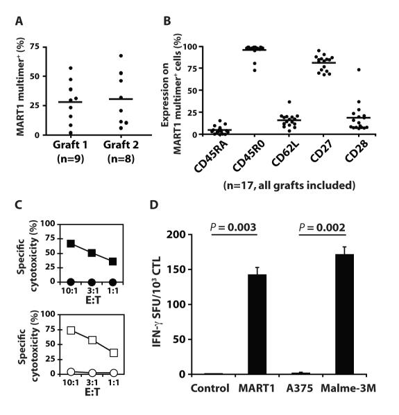
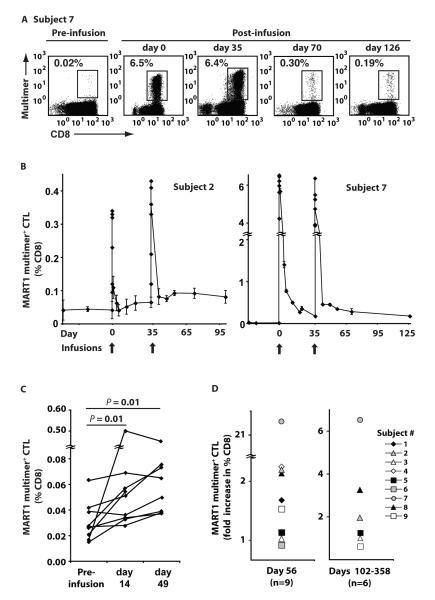
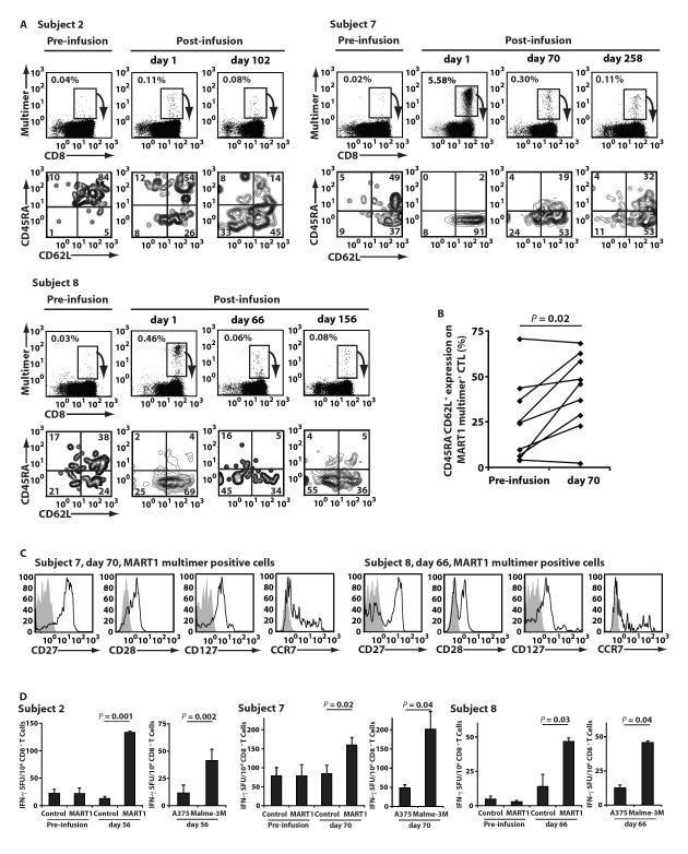
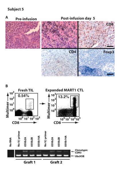
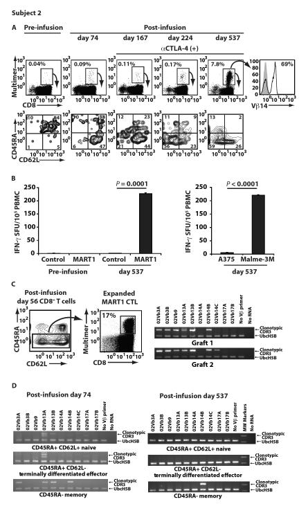
Although advanced-stage melanoma patients have a median survival of less than a year, adoptive T cell therapy can induce durable clinical responses in some patients. Successful adoptive T cell therapy to treat cancer requires engraftment of antitumor T lymphocytes that not only retain specificity and function in vivo but also display an intrinsic capacity to survive. To date, adoptively transferred antitumor CD8(+) T lymphocytes (CTLs) have had limited life spans unless the host has been manipulated. To generate CTLs that have an intrinsic capacity to persist in vivo, we developed a human artificial antigen-presenting cell system that can educate antitumor CTLs to acquire both a central memory and an effector memory phenotype as well as the capacity to survive in culture for prolonged periods of time. We examined whether antitumor CTLs generated using this system could function and persist in patients. We showed that MART1-specific CTLs, educated and expanded using our artificial antigen-presenting cell system, could survive for prolonged periods in advanced-stage melanoma patients without previous conditioning or cytokine treatment. Moreover, these CTLs trafficked to the tumor, mediated biological and clinical responses, and established antitumor immunologic memory. Therefore, this approach may broaden the availability of adoptive cell therapy to patients both alone and in combination with other therapeutic modalities.


Figures

Fig. 1
Fig. 1. Infused MART1-specific CTL grafts possessed a central memory and effector memory phenotype and effector function
Seventeen MART1-specific CTL grafts were generated and administered to 9 patients. (A) The MART1 multimer positive percentage of infused CTL grafts is shown separately for grafts 1 (n=9) and 2 (n=8) for all patients. Bars represent the mean values. (B) Percent expression for the indicated molecules on MART1 multimer+ CTL is shown for all grafts (n=17). (C and D) A representative example of functional assays for infused MART1-specific CTL grafts is shown (Subject 5). (C) Antigen-specific cytotoxicity was demonstrated for MART1 peptide pulsed T2 (■) versus control peptide pulsed T2 targets (•), and the HLA-A2+ MART1+ melanoma line, Malme-3M (□), versus the HLA-A2+ MART1 melanoma line, A375 (○). (D) IFN-γ ELISPOT showed antigen-specific IFN-γ secretion using peptide pulsed T2 cells and tumor cell line targets. Mean values ±SD of triplicates are shown.
Fig. 2
Fig. 2. Adoptive transfer induced sustained increases in the frequency of circulating MART1-specific CD8+ T cells
The frequency of MART1-specific T cells was determined by MART1 multimer staining of circulating CD8+ T cells pre- and post-infusion without any in vitro expansion. Note that patients received no other therapies such as CTLA-4 blockade during the indicated time periods. (A) Representative MART1 multimer staining for Subject 7 is shown. Day 0 and 35 analyses were performed on blood samples drawn 30 minutes post-infusion of CTL grafts. (B) The frequency of MART1-specific CD8+ T cells over the course of the clinical study is shown for Subjects 2 and 7. Mean values ±SD of quadruplicates are shown. (C) The frequency of MART1-specific CD8+ T cells for all patients is shown for time points pre-infusion and 14 days post-infusion. The average of three pre-infusion time points was used as the baseline. (D) A sustained increase in the frequency of MART1-specific T cells on day 56 was observed and is expressed as the ratio of multimer positive CD8+ T cells on day 56/pre-infusion baseline (left). Data for day 21 was included for Subject 1, who did not receive a second infusion. The ratio of multimer positive CD8+ T cells at later time points/pre-infusion baseline is shown (right). Post-infusion samples were obtained for Subjects 2, 3, 5, 7, 8, and 9 on days 102, 145, 134, 258, 358, and 133 respectively.
Fig. 3
Fig. 3. Adoptive transfer increased the number of MART1-specific T cells with a central memory phenotype and memory function
(A) The phenotype of circulating MART1-specific T cells was assessed for all 9 patients pre- and post-infusion. In the depicted examples (Subjects 2, 7, and 8), the CD45RA/CD62L phenotype of fresh, circulating MART1 multimer+ cells is shown below the multimer stain for each indicated time point. Note that patients received no other anti-melanoma therapies such as anti-CTLA-4 mAb treatment during the indicated periods. (B) The percentage of peripheral CD45RA CD62L+ MART1 multimer+ CTL is shown for all 9 patients pre- and post-infusion (P<0.05). Data for day 35 was included for Subject 1, who did not receive a second infusion. (C) The phenotype of MART1-specific CD8+ T cells for Subject 7, day 70, and Subject 8, day 66, was demonstrated. The phenotype of gated MART1 multimer staining cells is shown (open) with isotype control mAb staining (shaded). (D) Pre- and post-infusion MART1-specific recall responses are shown in IFN-γ ELISPOT assays for Subjects 2, 7, and 8. Without any prior expansion, freshly purified, peripheral CD8+ T cells were stimulated with T2 targets pulsed with MART1 or control peptide (left). T cells were also incubated with melanoma cell lines, A375 (HLA-A2+ MART1) and Malme-3M (HLA-A2+ MART1+) (right). Data shown represents mean values ±SD of triplicates.
Fig. 4
Fig. 4. Transferred CTL trafficked to sites of antigen expression and mediated anti-tumor responses
Pathologic analysis was performed to determine whether transferred T cells were able to traffic to sites of antigen expression. (A) Infiltration of tumor by lymphocytes was morphologically assessed pre-infusion and post-infusion for Subject 5. Immunohistochemical staining of the post-infusion biopsy for CD8, CD4 and Foxp3 is shown. Scale bar, 50 μm, upper right, applies to upper panels. Scale bar, 100 μm, lower right, applies to lower panels. (B) Without in vitro expansion, MART1-specific T cells were identified in fresh TIL by MART1 multimer staining. Three MART1-specific clonotypes derived from TIL were identified. The presence of all three MART1-specific T cell clonotypes in both CTL grafts was shown by RT-PCR.
Fig. 5
Fig. 5. Adoptively transferred CTL expanded in a patient treated with anti-CTLA-4 (ipilimumab) therapy
After two infusions of MART1 CTL, Subject 2 began anti-CTLA-4 mAb therapy on post-infusion day 103. (A) The frequency of peripheral MART1-specific T cells is shown pre- and post-infusion of CTL and after initiation of anti-CTLA-4 mAb therapy. The CD45RA/CD62L phenotype of multimer positive cells is shown below the multimer stain for each time point. TCR Vβ 14 staining of markedly expanded MART1 multimer+ T cells on day 537 is shown (right). (B) Without any prior in vitro expansion, MART1-specific recall responses on day 537, but not pre-infusion, were demonstrated with the IFN-γ ELISPOT assay using PBMC incubated with control or MART1 peptide (left). Fresh day 537 PBMC were also incubated with the melanoma cell lines, A375 (HLA-A2+ MART1) and Malme-3M (HLA-A2+ MART1+) (right). Data shown represents mean values ±SD of triplicates. (C) MART1-specific CTL were expanded from the memory fraction (CD45RA) of CD8+ T cells on day 56, prior to CTLA-4 blockade, to identify 10 MART1-specific clonotypes. The presence of seven of these 10 clones in CTL grafts 1 or 2, including Vβ 14 clones, 02Vb14B and 02Vb14C, is shown. (D) Identification of the Vβ 14 clone, 02Vb14B, exclusively in the CD45RA memory subpopulation only is shown on day 74, prior to CTLA-4 blockade (left), and on day 537, after CTLA-4 blockade (right).

References

    1. Hodi FS, O’Day SJ, McDermott DF, Weber RW, Sosman JA, Haanen JB, Gonzalez R, Robert C, Schadendorf D, Hassel JC, Akerley W, van den Eertwegh AJ, Lutzky J, Lorigan P, Vaubel JM, Linette GP, Hogg D, Ottensmeier CH, Lebbe C, Peschel C, Quirt I, Clark JI, Wolchok JD, Weber JS, Tian J, Yellin MJ, Nichol GM, Hoos A, Urba WJ. Improved survival with ipilimumab in patients with metastatic melanoma. N Engl J Med. 2010 - PMC - PubMed
    1. Dudley ME, Yang JC, Sherry R, Hughes MS, Royal R, Kammula U, Robbins PF, Huang J, Citrin DE, Leitman SF, Wunderlich J, Restifo NP, Thomasian A, Downey SG, Smith FO, Klapper J, Morton K, Laurencot C, White DE, Rosenberg SA. Adoptive cell therapy for patients with metastatic melanoma: Evaluation of intensive myeloablative chemoradiation preparative regimens. J Clin Oncol. 2008;26:5233–5239. - PMC - PubMed
    1. Berger C, Turtle CJ, Jensen MC, Riddell SR. Adoptive transfer of virus-specific and tumor-specific t cell immunity. Curr Opin Immunol. 2009;21:224–232. - PMC - PubMed
    1. Rosenberg SA, Dudley ME. Adoptive cell therapy for the treatment of patients with metastatic melanoma. Curr Opin Immunol. 2009;21:233–240. - PMC - PubMed
    1. Brenner MK, Heslop HE. Adoptive t cell therapy of cancer. Curr Opin Immunol. 2010;22:251–257. - PMC - PubMed

Publication types

MeSH terms