. 2011 Aug;36(9):1801-10.
doi: 10.1038/npp.2011.59. Epub 2011 Apr 27.

Cholecystokinin exerts an effect via the endocannabinoid system to inhibit GABAergic transmission in midbrain periaqueductal gray

Affiliations

Cholecystokinin exerts an effect via the endocannabinoid system to inhibit GABAergic transmission in midbrain periaqueductal gray

Vanessa A Mitchell et al. Neuropsychopharmacology. 2011 Aug.

Abstract

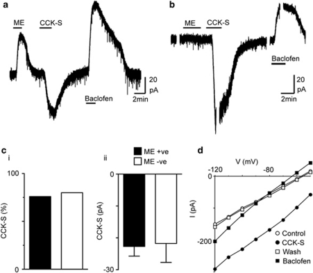
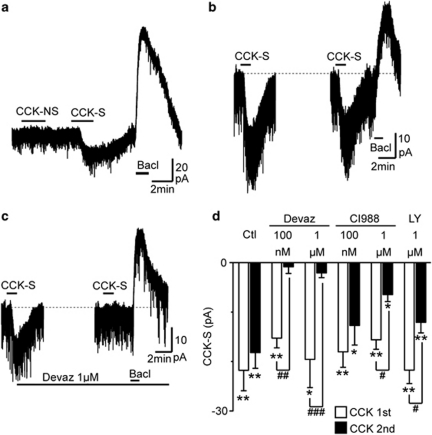
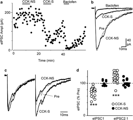
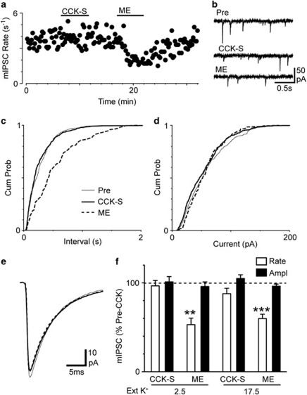
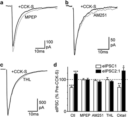
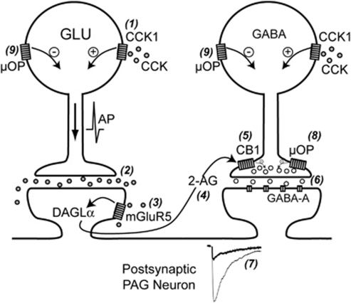
Cholecystokinin modulates pain and anxiety via its functions within brain regions such as the midbrain periaqueductal gray (PAG). The aim of this study was to examine the cellular actions of cholecystokinin on PAG neurons. Whole-cell patch clamp recordings were made from rat midbrain PAG slices in vitro to examine the postsynaptic effects of cholecystokinin and its effects on synaptic transmission. Sulfated cholecystokinin-(26-33) (CCK-S, 100-300 nM), but not non-sulfated cholecystokinin-(26-33) (CCK-NS, 100-300 nM) produced an inward current in a sub-population of opioid sensitive and insensitive PAG neurons, which did not reverse over a range of membrane potentials. The CCK-S-induced current was abolished by the CCK1 selective antagonist devazepide (100 nM), but not by the CCK2 selective antagonists CI988 (100 nM, 1 μM) and LY225910 (1 μM). CCK-S, but not CCK-NS produced a reduction in the amplitude of evoked GABA(A)-mediated inhibitory postsynaptic currents (IPSCs) and an increase in the evoked IPSC paired-pulse ratio. By contrast, CCK-S had little effect on the rate and amplitude of TTX-resistant miniature IPSCs under basal conditions and when external K(+) was elevated. The CCK-S-induced inhibition of evoked IPSCs was abolished by the cannabinoid CB1 receptor antagonist AM251 (3 μM), the mGluR5 antagonist MPEP (10 μM) and the 1, 2-diacylglycerol lipase (DAGLα) inhibitor tetrahydrolipstatin (10 μM). In addition, CCK-S produced an increase in the rate of spontaneous non-NMDA-mediated, TTX-dependent excitatory postsynaptic currents (EPSCs). These results suggest that cholecystokinin produces direct neuronal depolarisation via CCK1 receptors and inhibits GABAergic synaptic transmission via action potential-dependent release of glutamate and mGluR5-induced endocannabinoid signaling. Thus, cholecystokinin has cellular actions within the PAG that can both oppose and reinforce opioid and cannabinoid modulation of pain and anxiety within this brain structure.


Figures

Figure 1
Figure 1
Cholecystokinin produces an inward current in sub-populations of μ-opioid sensitive and insensitive neurons. Current traces of opioid (a) sensitive and (b) insensitive PAG neurons during superfusion of met-enkephalin (ME, 10 μM), sulfated cholecystokinin-(26–33) (CCK-S, 300 nM) and baclofen (10 μM). (c) Bar charts depicting the (i) percentage of neurons that responded to CCK-S with an inward current, and (ii) the average inward current in CCK-S-responding neurons for ME responding and non-responding neurons (ME +ve and −ve). (d) Current–voltage relationship for the neuron in (b) before (control), during (CCK-S), and after washout (wash) of CCK-S and then during baclofen. Membrane currents were evoked by voltage steps in 10 mV increments from –50 mV to –120 mV (250 ms duration). Current traces in (a and b) are from different neurons.
Figure 2
Figure 2
The postsynaptic effects of cholecystokinin are largely mediated by CCK1 receptors. (a) Current trace of a neuron during superfusion of non-sulfated cholecystokinin-(26–33) (CCK-NS, 100 nM), sulfated cholecystokinin-(26–33) (CCK-S, 100 nM), and baclofen (Bacl, 10 μM). (b, c) Current traces of two neurons during repeated application of CCK-S (100 nM) at a 10 min interval, and then baclofen (10 μM). In (c) devazepide (1 μM) was added after the first washout of CCK-S. (d) Bar chart showing the inward currents produced during two consecutive applications of 100 nM CCK-S (CCK 1st and 2nd), in which either no antagonist (Ctl, Control), devazepide (Devaz, 100 nM, 1 μM), CI988 (100 nM, 1 μM), or LY225910 (LY, 1 μM) was applied continuously after washout of the first application of CCK-S. In (d) *p<0.05 and **p<0.01 (t-tests for 1st and 2nd CCK-S currents individually), #p<0.05, ##p<0.01 and ###p<0.001 for post-hoc comparisons between 1st and 2nd CCK-S currents for individual treatment groups with two-way ANOVA. Current traces in (a–c) are from different neurons.
Figure 3
Figure 3
Cholecystokinin inhibits evoked inhibitory postsynaptic currents (IPSCs). (a) Time course of evoked IPSC amplitude (eIPSC Ampl) during application of non-sulfated cholecystokinin-(26–33) (CCK-NS, 300 nM), sulfated cholecystokinin-(26–33) (CCK-S, 300 nM), and then baclofen (10 μM). (b) Averaged evoked IPSCs before (Pre), and during application of CCK-NS, CCK-S, and baclofen. (c) Averaged evoked IPSCs in response to identical paired stimuli (inter-stimulus interval=80 ms) before (Pre) and during CCK-NS and CCK-S, with IPSC1 normalized to demonstrate relative facilitation of IPSC2 during superfusion of CCK-S. (d) Scatter plot showing the amplitude of the first evoked IPSC (eIPSC1) and the ratio of evoked IPSC2/IPSC1 (eIPSC2 : 1) in the presence of CCK-NS and CCK-S expressed as a percentage of the pre-CCK-NS and CCK-S values, respectively. In (d) ***p<0.001. Traces in (a–c) are from the same neuron. In (b–c) stimulus artifacts have been blanked for clarity.
Figure 4
Figure 4
Effect of cholecystokinin on miniature IPSCs. (a) Time course of miniature inhibitory postsynaptic current (IPSC) (mIPSC) rate during superfusion of sulfated cholecystokinin-(26–33) (CCK-S, 300 nM) and met-enkephalin (ME, 10 μM) in normal artificial cerebrospinal fluid (ACSF). (b) Raw current traces of miniature IPSCs before (Pre) and during superfusion of CCK-S and ME. Cumulative probability distribution plots of miniature IPSC, (c) inter-event interval, and (d) amplitude, before and during superfusion of CCK-S and ME. (e) Averaged traces of miniature IPSCs before and during superfusion of CCK-S and ME, for the corresponding epochs used in (c) and (d). (f) Bar chart of the mean rate and amplitude of miniature IPSCs in the presence of CCK-S and ME, expressed as a percentage of the pre-drug value, in normal ACSF (K+=2.5 mM) and in elevated external K+ (17.5 mM). In (f) **p<0.01 and ***p<0.001. Traces in (a–e) are from the same neuron.
Figure 5
Figure 5
Cholecystokinin inhibition of evoked inhibitory postsynaptic currents (IPSCs) is mediated by mGluR5-induced endocannabinoid signaling. Traces of averaged evoked IPSCs before and after addition of sulfated cholecystokinin-(26–33) (CCK-S, 300 nM) in PAG neurons from slices, which were pre-incubated in either (a) the mGluR5 antagonist MPEP (5 μM), (b) the cannabinoid CB1 antagonist AM251 (1 μM), or (c) the DAG lipase inhibitor tetrahydrolipstatin (THL, 10 μM). (d) Bar chart showing the effect of CCK-S (300 nM) on the first evoked IPSC (eIPSC1) and the ratio of evoked IPSC2/IPSC1 (eIPSC2 : 1), in control untreated slices (Ctl) and slices pre-incubated in either MPEP, AM251, THL, or a cocktail of NK1 (L732138 10 μM), NTS1/2 (SR142948 300 nM) and mAChR (atropine 1 μM) antagonists (Cktail). The data in (d) is the mean eIPSC amplitude and eIPSC2 : 1 ratio in the presence of CCK-S expressed as a percentage of the pre-CCK-S value. In (d) *p<0.05, **p<0.01 and ***p<0.001 (comparing values before and during addition of CCK-S). Traces in (a–c) are from different neurons, and stimulus artifacts have been blanked for clarity.
Figure 6
Figure 6
Proposed model for CCK-induced suppression of GABAergic transmission in PAG. Cholecystokinin (CCK) activates postsynaptic CCK1 (and possibly CCK2) receptors located on glutamatergic (GLU) neurons (1) to elicit action potential (AP)-driven glutamate release (2). This endogenous glutamate then activates postsynaptic mGluR5 receptors (3), which causes the production of the endocannabinoid 2-arachidonoylglycerol (2-AG) via the enzyme 1,2-diacylglycerol lipase (DAGLα) (4). 2-AG leaves the postsynaptic neuron, exerts an effect as a retrograde messenger to activate presynaptic cannabinoid CB1 receptors located on the terminals of GABAergic neurons (5) and reduces GABA release (6). The subsequent inhibition of GABAergic synaptic transmission (7) leads to disinhibition (excitation) of the postsynaptic neuron. It might be noted that the CCK-induced endocannabinoid inhibition of GABAergic synaptic transmission will be enhanced by agonists exerting an effect at presynaptic μ-opioid receptors (8). By contrast, neuronal excitation produced by postsynaptic CCK1 receptor activation will oppose postsynaptic μ-opioid receptor-mediated inhibition (9).

References

    1. Bertoglio LJ, Zangrossi H., Jr Involvement of dorsolateral periaqueductal gray cholecystokinin-2 receptors in the regulation of a panic-related behavior in rats. Brain Res. 2005;1059:46–51. - PubMed
    1. Bisogno T, Howell F, Williams G, Minassi A, Cascio MG, Ligresti A, et al. Cloning of the first sn1-DAG lipases points to the spatial and temporal regulation of endocannabinoid signaling in the brain. J Cell Biol. 2003;163:463–468. - PMC - PubMed
    1. Brack KE, Jeffery SM, Lovick TA. Cardiovascular and respiratory responses to a panicogenic agent in anaesthetised female Wistar rats at different stages of the oestrous cycle. Eur J Neurosci. 2006;23:3309–3318. - PubMed
    1. Brack KE, Lovick TA. Neuronal excitability in the periaqueductal grey matter during the estrous cycle in female Wistar rats. Neuroscience. 2007;144:325–335. - PubMed
    1. Carlberg M, Gundlach AL, Mercer LD, Beart PM. Autoradiographic localization of cholecystokinin A and B receptors in rat brain using [125I]d-Tyr25 (Nle28,31)-CCK 25–33S. Eur J Neurosci. 1992;4:563–573. - PubMed

Publication types

MeSH terms