. 2011 Apr 19;6(4):e19124.
doi: 10.1371/journal.pone.0019124.

The telomere binding protein TRF2 induces chromatin compaction

Affiliations

The telomere binding protein TRF2 induces chromatin compaction

Asmaa M Baker et al. PLoS One. .

Abstract

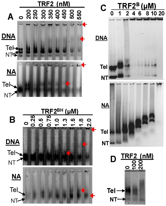
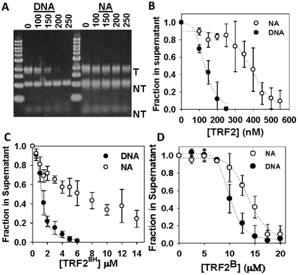
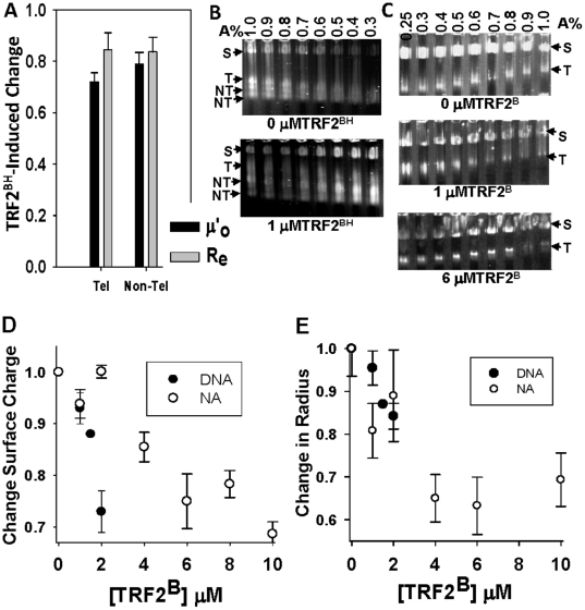
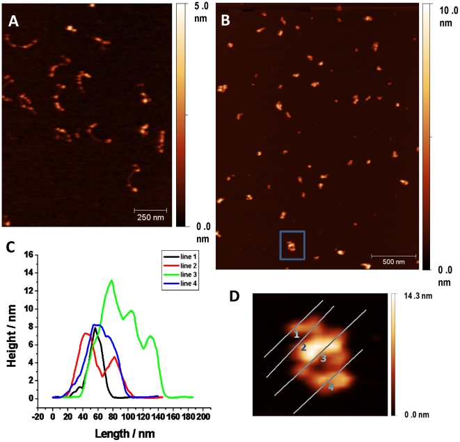
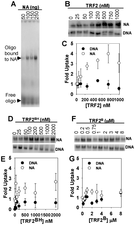
Mammalian telomeres are specialized chromatin structures that require the telomere binding protein, TRF2, for maintaining chromosome stability. In addition to its ability to modulate DNA repair activities, TRF2 also has direct effects on DNA structure and topology. Given that mammalian telomeric chromatin includes nucleosomes, we investigated the effect of this protein on chromatin structure. TRF2 bound to reconstituted telomeric nucleosomal fibers through both its basic N-terminus and its C-terminal DNA binding domain. Analytical agarose gel electrophoresis (AAGE) studies showed that TRF2 promoted the folding of nucleosomal arrays into more compact structures by neutralizing negative surface charge. A construct containing the N-terminal and TRFH domains together altered the charge and radius of nucleosomal arrays similarly to full-length TRF2 suggesting that TRF2-driven changes in global chromatin structure were largely due to these regions. However, the most compact chromatin structures were induced by the isolated basic N-terminal region, as judged by both AAGE and atomic force microscopy. Although the N-terminal region condensed nucleosomal array fibers, the TRFH domain, known to alter DNA topology, was required for stimulation of a strand invasion-like reaction with nucleosomal arrays. Optimal strand invasion also required the C-terminal DNA binding domain. Furthermore, the reaction was not stimulated on linear histone-free DNA. Our data suggest that nucleosomal chromatin has the ability to facilitate this activity of TRF2 which is thought to be involved in stabilizing looped telomere structures.


Conflict of interest statement

Competing Interests: The authors have declared that no competing interests exist.

Figures

Figure 1
Figure 1. TRF2 binds to telomeric DNA (DNA) and nucleosomal array fibers (NA).
TRF2 (A) or TRF2BH (B) binding to substrates detected by electrophoresis on 0.3% agarose gels or 0.6% agarose gels to detect binding of TRF2B (C). DNA and nucleosomal arrays pertain to pRST5 digested to obtain a 2 kb fragment containing the 580 bp telomeric DNA (Tel) with a 1 kb and smaller fragments being non-telomeric (NT). 0.6% agarose gel to detect binding of TRF2 to nucleosomal arrays derived from digestion of with SfaNI/PvuII/BspHI (D).The 0.3% agarose lanes in (A) and (B) were formed using a multi-gel apparatus as described in Materials and Methods. Red arrows point to mobility shifts produced by TRF2 or TRF2BH complexes.
Figure 2
Figure 2. TRF2 stimulates self-association of DNA and nucleosomal arrays.
Differential centrifugation assay as described in Materials and Methods. 1% agarose gel of samples with indicated amounts of TRF2 in nM where “T” refers to telomeric and “NT” refers to non-telomeric fragments (A). Quantification of experiments with TRF2 (B) TRF2BH (C) and TRF2B (D). Each data point represents the mean ± 1 SD from 3 separate experiments.
Figure 3
Figure 3. TRF2-dependent changes in surface charge density (µ'o) and effective radius (Re from dilute gels) of nucleosomal fibers determined by analytical agarose gel electrophoresis (AAGE).
Multi-gels of telomeric nucleosomal array fibers (NA) in the absence (A) or presence (B) of 200 nM TRF2 prepared and subjected to electrophoresis according to Materials and Methods. “S” refers to carboxylate-coated microsphere standards (35 nm radius). “T” refers to the telomeric fragments liberated by SfaNI/PvuII/BspHI digestion of pRST5 and “NT” refers to the non-telomeric DNA fragments. TRF2-induced change in surface charge density (µ'o) and effective radius (Re) of nucleosomal arrays derived from the telomeric (Tel) or non-telomeric (non-Tel) fragments (C). The µ'o (black bars) or Re (grey bars) of NA in the presence of 200 nM TRF2 was normalized to 0 nM TRF2. Bars represent the mean ±1 SD from 3 separate experiments. The data were derived from multi-gels of 0.25–1% agarose concentrations while the Re bars represent the average from 0.25–0.6% agarose concentrations according to Materials and Methods.
Figure 4
Figure 4. The role of the TRF2 basic N-terminus alone (TRF2B) or with the TRFH domain (TRF2BH) in TRF2-dependent negative charge reduction and compaction of nucleosomal arrays (NA).
TRF2BH-induced change in surface charge density (µ'o) and effective radius (Re) of nucleosomal arrays derived from the telomeric (Tel) or largest non-telomeric (non-Tel) fragment (A). Bars represent the mean ±1 SD of 3 multi-gel experiments. The µ'o (black bars) or Re (grey bars) of NA in the presence of 1 µM TRF2BH was normalized to 0 µM TRF2BH. Multi-gels of telomeric nucleosomal array fibers (NA) in the absence or presence of 1 µM TRF2BH (B) prepared and subjected to electrophoresis according to Materials and Methods. “S” refers to carboxylate-coated microsphere standards (35 nm radius). “T” refers to the telomeric fragments liberated by SfaNI/PvuII/BspHI digestion of pRST5 and “NT” refers to the non-telomeric DNA fragments. Multi-gels of telomeric nucleosomal array fibers (NA) in the presence of indicated amounts of TRF2B (C). TRF2B-induced changes in surface charge density (µ'o). (D) and effective radius (Re from dilute gels) (E) of DNA and nucleosomal arrays (NA). The µ'o or Re for each TRF2B concentration was normalized to 0 µM TRF2B. Each data point represents the mean ±1 SD of 3–4 multi-gel experiments.
Figure 5
Figure 5. Atomic force microscopy of TRF2B-nucleosomal array complexes.
Nucleosomal array fibers (reconstituted with 1∶1 histone:DNA mass ratio) in the absence of TRF2B (A). Nucleosomal arrays with 4 µM TRF2B (B). An example of height measurements (C) of regions indicated by lines drawn on the fiber (D) expanded from in the boxed region in (B). Samples were prepared and analyzed according to Materials and Methods.
Figure 6
Figure 6. The effect of full-length TRF2, TRF2BH, and TRF2B on the insertion of a 5′-[32P]-labeled, single-stranded oligonucleotide, (dTTAGGG)7 (T7), into nucleosomal arrays and DNA (20 ng/µl).
Samples were incubated with indicated amounts TRF2 or its truncated mutants and processed according to Materials and Methods. Agarose gel showing insertion of T7 (Free oligo) into increasing amounts of nucleosomal arrays (Oligo bound to NA), as indicated (A). The section of agarose gels showing T7 inserted into nucleosomal arrays (NA) or linear DNA (DNA) with increasing TRF2 or truncation mutants as indicated (B, D and F). Quantification of corresponding gels above where uptake was normalized to 0 nM TRF2 or truncation mutants (C, E and G). Each data point represents the mean ±1 SD from 3–4 separate experiments.

Similar articles

Cited by

References

    1. Falk M, Lukasova E, Kozubek S. Higher-order chromatin structure in DSB induction, repair and misrepair. Mutat Res. 2010;704:88–100. - PubMed
    1. Hansen JC. Conformational dynamics of the chromatin fiber in solution: determinants, mechanisms, and functions. Annu Rev Biophys Biomol Struct. 2002;31:361–392. - PubMed
    1. Horn PJ, Peterson CL. Molecular biology. Chromatin higher order folding—wrapping up transcription. Science. 2002;297:1824–1827. - PubMed
    1. Luger K, Hansen JC. Nucleosome and chromatin fiber dynamics. Curr Opin Struct Biol. 2005;15:188–196. - PubMed
    1. Lu X, Klonoski JM, Resch MG, Hansen JC. In vitro chromatin self-association and its relevance to genome architecture. Biochem Cell Biol. 2006;84:411–417. - PubMed

Publication types