. 2011 Apr 22;6(4):e17887.
doi: 10.1371/journal.pone.0017887.

High affinity antigen recognition of the dual specific variants of herceptin is entropy-driven in spite of structural plasticity

Affiliations

High affinity antigen recognition of the dual specific variants of herceptin is entropy-driven in spite of structural plasticity

Jenny Bostrom et al. PLoS One. .

Abstract

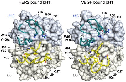
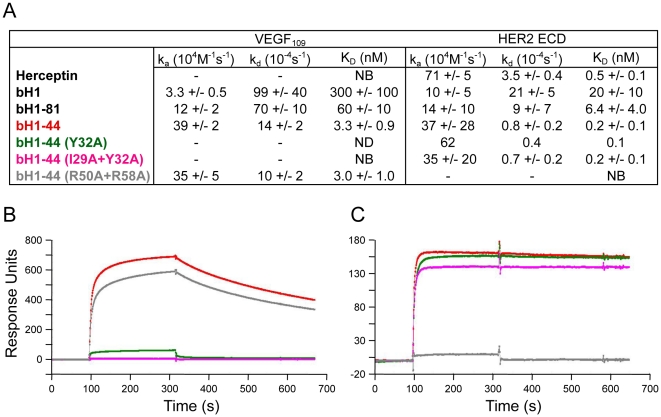
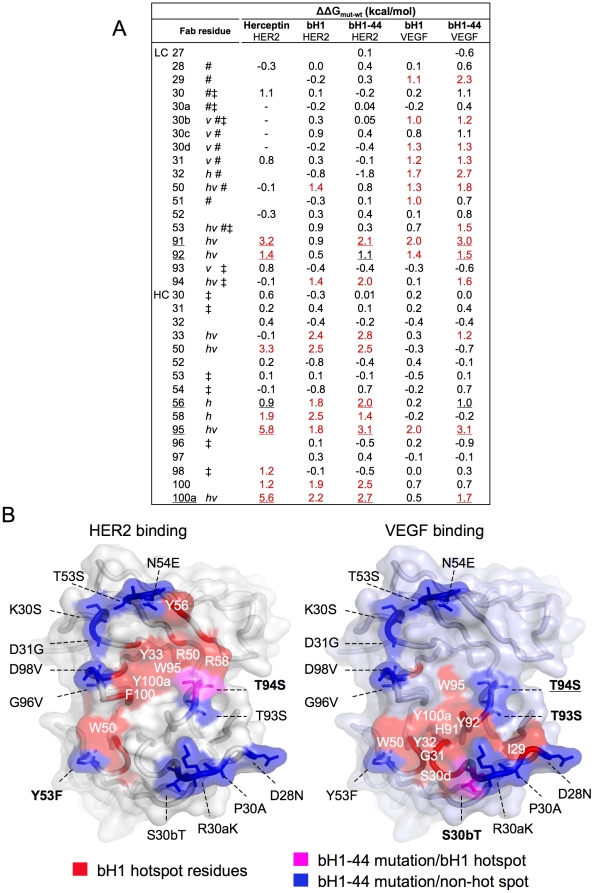
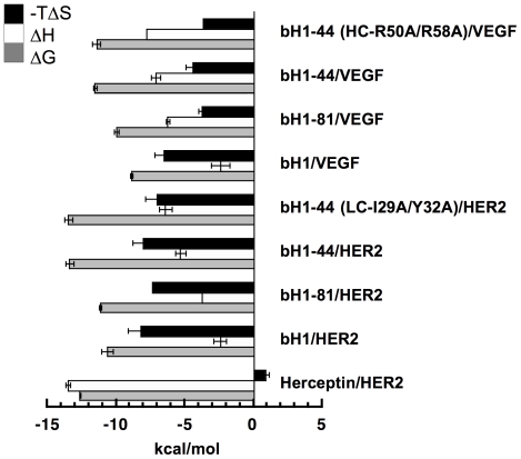
The antigen-binding site of Herceptin, an anti-human Epidermal Growth Factor Receptor 2 (HER2) antibody, was engineered to add a second specificity toward Vascular Endothelial Growth Factor (VEGF) to create a high affinity two-in-one antibody bH1. Crystal structures of bH1 in complex with either antigen showed that, in comparison to Herceptin, this antibody exhibited greater conformational variability, also called "structural plasticity". Here, we analyzed the biophysical and thermodynamic properties of the dual specific variants of Herceptin to understand how a single antibody binds two unrelated protein antigens. We showed that while bH1 and the affinity-improved bH1-44, in particular, maintained many properties of Herceptin including binding affinity, kinetics and the use of residues for antigen recognition, they differed in the binding thermodynamics. The interactions of bH1 and its variants with both antigens were characterized by large favorable entropy changes whereas the Herceptin/HER2 interaction involved a large favorable enthalpy change. By dissecting the total entropy change and the energy barrier for dual interaction, we determined that the significant structural plasticity of the bH1 antibodies demanded by the dual specificity did not translate into the expected increase of entropic penalty relative to Herceptin. Clearly, dual antigen recognition of the Herceptin variants involves divergent antibody conformations of nearly equivalent energetic states. Hence, increasing the structural plasticity of an antigen-binding site without increasing the entropic cost may play a role for antibodies to evolve multi-specificity. Our report represents the first comprehensive biophysical analysis of a high affinity dual specific antibody binding two unrelated protein antigens, furthering our understanding of the thermodynamics that drive the vast antigen recognition capacity of the antibody repertoire.


Conflict of interest statement

Competing Interests: All authors are or were paid employees of Genentech, Inc., which is a member of Roche Group, and all authors own shares of Roche stocks. J.B. and G.F. are inventors of a patent application, “Multispecific Antibodies” (Pub. App. No. 20080069820), which is related to this work. The authors confirm here that these conflicts do not alter their adherence to all the PLoS ONE policies on sharing data and materials described in their report that are reasonably requested by others for the purpose of academic, non-commercial research.

Figures

Figure 1
Figure 1. The distinct CDR conformations of bH1.
The three HC CDRs (cyan) and three LC CDRs (yellow) of bH1 in complex with HER2 (PDB code 3BE1) or VEGF (PDB code 3BDY) are shown in cartoon representation with the rest of bH1 structure as surface. Selected residues are shown in stick representation: LC-I29, Y32 and I30c are specifically important for bH1-44/VEGF binding and HC-R50 and R58 are specifically important for bH1-44/HER2 binding whereas LC-H91 and Y92, and HC-Y56, W95 and Y100a (in bold) are residues important for the bH1-44 interaction with both antigens as well as the Herceptin interaction with HER2. Note the highly distinct conformations of CDR-L1, the adjustment of the side chains of highlighted residues, and the side chains of LC-I30c and LC-Y32 that alternatively occupy the nearby cavity (*) in HER2 bound bH1 or VEGF bound bH1, respectively. See Figure S1 movie (Movie S1) morphing the two structures, which highlights the extent of the conformational adjustment for dual interaction.
Figure 2
Figure 2. Antigen-binding affinity and kinetic of the bH1 variants and Herceptin.
(A) kon and koff and dissociation constant (KD) of Fabs binding to immobilized VEGF or HER2 are determined by SPR measurements (See Methods). The errors represent the standard deviations based on at least three independent experiments. bH1-81 was measured only once. Hence no error estimation is available. NB = No binding detected. ND = Not determined as the interaction was too weak to assess. The representative binding responses over time of 0.5 µM Fab bH1-44 (red), bH1-44 (LC-Y32) (green), bH1-44(LC-I29A+Y32A) (magenta) and bH1-44(HC-R50A+R58A) (gray) to immobilized VEGF109 (B) or HER2-ECD (C).
Figure 3
Figure 3. Mapping of the evolved dual specific interactions.
The changes in binding free energy (ΔΔGmut-wt) when a Fab residue is mutated to alanine (based on data from Kelley et. al. Biochemistry, 1993, Bostrom et. al., Science, 2009, and the current study) are shown (A). Amino acids are numbered as in Table 1 and denoted with h or v if making structural contact with HER2 or VEGF (within 4.5 Å), respectively, # if mutated from Herceptin to bH1, and ‡ if mutated from bH1 to bH1-44. Underlined residues are energetically important (ΔΔGmut-wt greater than ∼1 kcal/mol, also underlined) for Herceptin/HER2, bH1-44/HER2 and bH1-44/VEGF interactions indicating importance for all three interactions. Hotspot residues highlighted in red are those with ΔΔGmut-wt greater than ∼10% of the total binding free energy of each interaction. “-” denotes no residue in Herceptin at the position. Values not determined are left blank. (B) bH1-44 mutations (from bH1) are mapped to locate predominantly outside of functional hotspots and structural contact for HER2 binding (left) or VEGF binding (right) on the surface of the bH1 paratope modeled with side chains of bH-44. The residues are colored red if they are bH1 hotspot residues for HER2 or VEGF binding, blue if the residues are mutated from bH1 to bH1-44 and not part of bH1 hotspot for each respective interaction, or magenta if the mutated residues are part of bH1 hotspot. A mutation is bolded if the residue is a structural contact site in the respective bH1 interface or underlined if the mutated residue gains significant functional importance.
Figure 4
Figure 4. Thermodynamic profiles of the bH1 variants and Herceptin.
The entropic component (−TΔS) and enthalpic component (ΔH) of the binding free energy (ΔG) measured at 30°C in phosphate buffer (pH 7.4) are shown in kcal/mol. ΔG was derived from the dissociation constant (KD) measured with SPR (See Figure 2) (ΔG = RTlnKD), and ΔH was measured using ITC. −TΔS was calculated from the ΔG and ΔH according to −TΔS = ΔG−ΔH. Error bars of ΔG and ΔH represent standard deviation of three independent measurements (exception: the enthalpies of the bH1-81/HER2 and bH1-44(R50A/R58A)/VEGF interactions were measured only once, thus no error bar), and the errors of −TΔS are combined errors of ΔG and ΔH.

References

    1. Reichert JM, Rosensweig CJ, Faden LB, Dewitz MC. Monoclonal antibody successes in the clinic. Nat Biotechnol. 2005;23:1073–1078. - PubMed
    1. Notkins AL. Polyreactivity of antibody molecules. Trends Immunol. 2004;25:174–179. - PubMed
    1. Foote J. Immunology. Isomeric antibodies. Science. 2003;299:1327–1328. - PubMed
    1. James LC, Roversi P, Tawfik DS. Antibody multispecificity mediated by conformational diversity. Science. 2003;299:1362–1367. - PubMed
    1. Mouquet H, Scheid JF, Zoller MJ, Krogsgaard M, Ott RG, et al. Polyreactivity increases the apparent affinity of anti-HIV antibodies by heteroligation. Nature. 2010;467:591–595. - PMC - PubMed

Publication types

MeSH terms