

Adverse events post smallpox-vaccination: insights from tail scarification infection in mice with Vaccinia virus
- PMID: 21526210
- PMCID: PMC3078145
- DOI: 10.1371/journal.pone.0018924
Adverse events post smallpox-vaccination: insights from tail scarification infection in mice with Vaccinia virus
Abstract
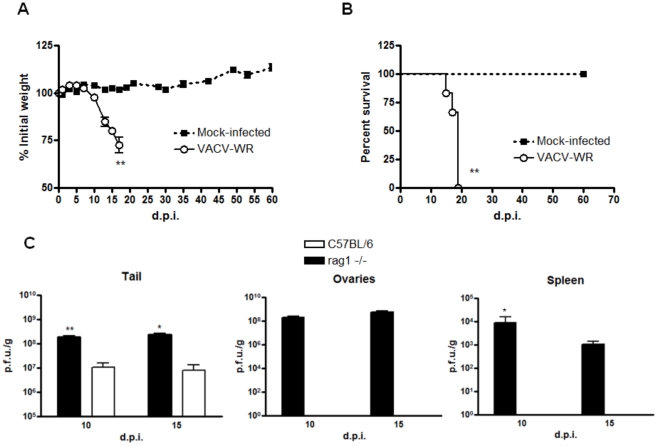
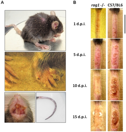
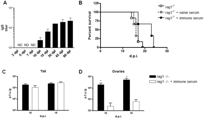
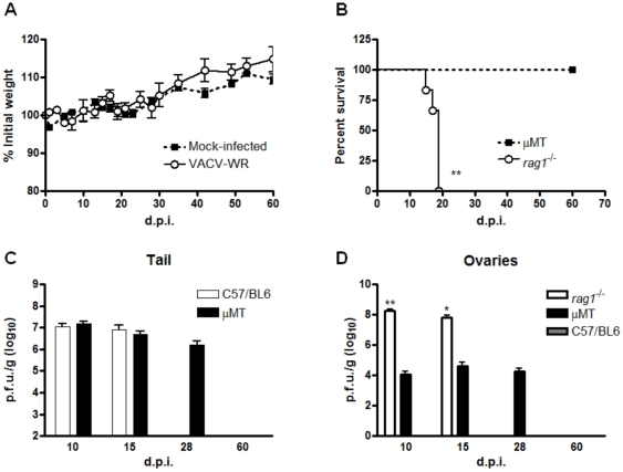
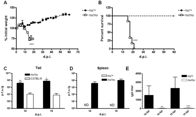
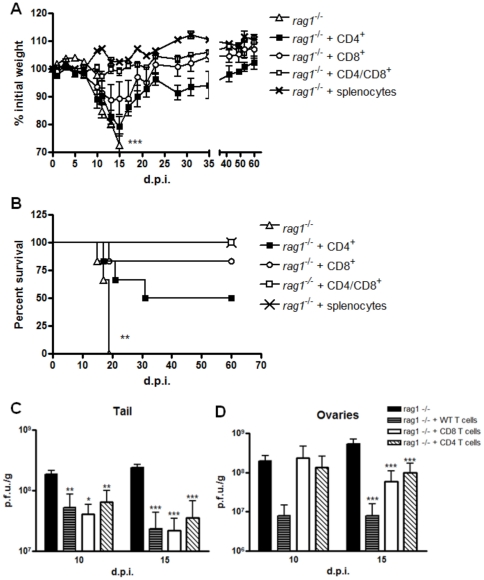
Adverse events upon smallpox vaccination with fully-replicative strains of Vaccinia virus (VACV) comprise an array of clinical manifestations that occur primarily in immunocompromised patients leading to significant host morbidity/mortality. The expansion of immune-suppressed populations and the possible release of Variola virus as a bioterrorist act have given rise to concerns over vaccination complications should more widespread vaccination be reinitiated. Our goal was to evaluate the components of the host immune system that are sufficient to prevent morbidity/mortality in a murine model of tail scarification, which mimics immunological and clinical features of smallpox vaccination in humans. Infection of C57BL/6 wild-type mice led to a strictly localized infection, with complete viral clearance by day 28 p.i. On the other hand, infection of T and B-cell deficient mice (Rag1(-/-)) produced a severe disease, with uncontrolled viral replication at the inoculation site and dissemination to internal organs. Infection of B-cell deficient animals (µMT) produced no mortality. However, viral clearance in µMT animals was delayed compared to WT animals, with detectable viral titers in tail and internal organs late in infection. Treatment of Rag1(-/-) with rabbit hyperimmune anti-vaccinia serum had a subtle effect on the morbidity/mortality of this strain, but it was effective in reduce viral titers in ovaries. Finally, NUDE athymic mice showed a similar outcome of infection as Rag1(-/-), and passive transfer of WT T cells to Rag1(-/-) animals proved fully effective in preventing morbidity/mortality. These results strongly suggest that both T and B cells are important in the immune response to primary VACV infection in mice, and that T-cells are required to control the infection at the inoculation site and providing help for B-cells to produce antibodies, which help to prevent viral dissemination. These insights might prove helpful to better identify individuals with higher risk of complications after infection with poxvirus.
Conflict of interest statement
Figures

Similar articles
-
Both CD8+ and CD4+ T Cells Contribute to Corneal Clouding and Viral Clearance following Vaccinia Virus Infection in C57BL/6 Mice.J Virol. 2016 Jun 24;90(14):6557-6572. doi: 10.1128/JVI.00570-16. Print 2016 Jul 15. J Virol. 2016. PMID: 27170749 Free PMC article.
-
Postchallenge administration of brincidofovir protects healthy and immune-deficient mice reconstituted with limited numbers of T cells from lethal challenge with IHD-J-Luc vaccinia virus.J Virol. 2015 Mar;89(6):3295-307. doi: 10.1128/JVI.03340-14. Epub 2015 Jan 14. J Virol. 2015. PMID: 25589648 Free PMC article.
-
Smallpox vaccine, ACAM2000: Sites and duration of viral shedding and effect of povidone iodine on scarification site shedding and immune response.Vaccine. 2015 Jun 12;33(26):2990-6. doi: 10.1016/j.vaccine.2015.04.062. Epub 2015 Apr 27. Vaccine. 2015. PMID: 25930115 Clinical Trial.
-
Pathogenesis and potential antiviral therapy of complications of smallpox vaccination.Antiviral Res. 2003 Apr;58(2):101-14. doi: 10.1016/s0166-3542(03)00008-1. Antiviral Res. 2003. PMID: 12742570 Review.
-
The immunology of smallpox vaccines.Curr Opin Immunol. 2009 Jun;21(3):314-20. doi: 10.1016/j.coi.2009.04.004. Epub 2009 Jun 11. Curr Opin Immunol. 2009. PMID: 19524427 Free PMC article. Review.
Cited by
-
Vaccinia Virus Vectors Targeting Peptides for MHC Class II Presentation to CD4+ T Cells.Immunohorizons. 2020 Jan 2;4(1):1-13. doi: 10.4049/immunohorizons.1900070. Immunohorizons. 2020. PMID: 31896555 Free PMC article.
-
Modulating Vaccinia Virus Immunomodulators to Improve Immunological Memory.Viruses. 2018 Feb 28;10(3):101. doi: 10.3390/v10030101. Viruses. 2018. PMID: 29495547 Free PMC article. Review.
-
Next-gen novel nanocage-based multivalent vaccine candidate to tackle the rising menace of Mpox.NPJ Vaccines. 2025 Jun 6;10(1):117. doi: 10.1038/s41541-025-01174-1. NPJ Vaccines. 2025. PMID: 40480973 Free PMC article.
-
Lamin A/C augments Th1 differentiation and response against vaccinia virus and Leishmania major.Cell Death Dis. 2018 Jan 8;9(1):9. doi: 10.1038/s41419-017-0007-6. Cell Death Dis. 2018. PMID: 29311549 Free PMC article.
-
Longitudinal Virological and Immunological Profile in a Case of Human Monkeypox Infection.Open Forum Infect Dis. 2022 Nov 1;9(12):ofac569. doi: 10.1093/ofid/ofac569. eCollection 2022 Dec. Open Forum Infect Dis. 2022. PMID: 36474633 Free PMC article.
References
-
- Moss B. Poxviridae: The viruses and their replication. 2001. Fundamental Virology. Forth Edition ed: Lippincott Williams & Wilkins.
-
- Fenner FHD, Arita I, Jezek Z, Ladnyi I. Geneva: World Health Organization; 1988. Smallpox and its eradication.
-
- Bray M, Wright ME. Progressive vaccinia. Clin Infect Dis. 2003;36:766–774. - PubMed
-
- Edghill-Smith Y, Golding H, Manischewitz J, King LR, Scott D, et al. Smallpox vaccine-induced antibodies are necessary and sufficient for protection against monkeypox virus. Nat Med. 2005;11:740–747. - PubMed
-
- Saito T, Fujii T, Kanatani Y, Saijo M, Morikawa S, et al. Clinical and immunological response to attenuated tissue-cultured smallpox vaccine LC16m8. JAMA. 2009;301:1025–1033. - PubMed
Publication types
MeSH terms
Substances
LinkOut - more resources
Full Text Sources
Medical
Molecular Biology Databases
