

Time course of collagen peak in bile duct-ligated rats
- PMID: 21527001
- PMCID: PMC3117813
- DOI: 10.1186/1471-230X-11-45
Time course of collagen peak in bile duct-ligated rats
Abstract
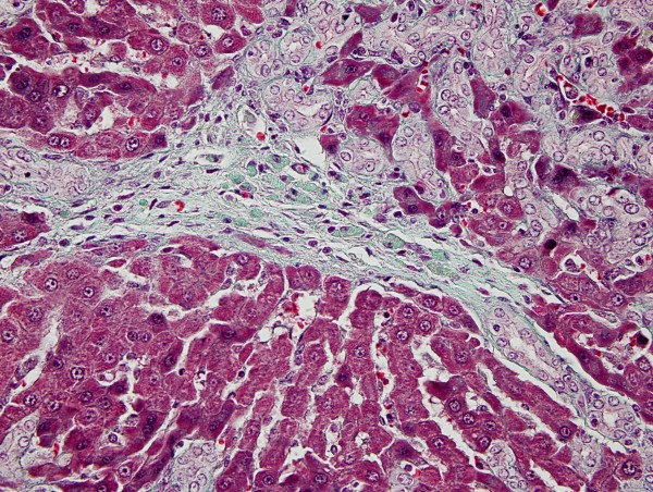
Background: One of the most useful experimental fibrogenesis models is the "bile duct-ligated rats". Our aim was to investigate the quantitative hepatic collagen content by two different methods during the different stages of hepatic fibrosis in bile duct-ligated rats on a weekly basis. We questioned whether the 1-wk or 4-wk bile duct-ligated model is suitable in animal fibrogenesis trials.
Methods: Of the 53 male Wistar rats, 8 (Group 0) were used as a healthy control group. Bile duct ligation (BDL) had been performed in the rest. Bile duct-ligated rates were sacrificed 7 days later in group 1 (10 rats), 14 days later in group 2 (9 rats), 21 days later in group 3(9 rats) and 28 days later in group 4 (9 rats). Eight rats underwent sham-operation (Sham). Hepatic collagen measurements as well as serum levels of liver enzymes and function tests were all analysed.
Results: The peak level of collagen was observed biochemically and histomorphometricly at the end of third week (P < 0.001 and P < 0.05). Suprisingly, collagen levels had decreased with the course of time such as at the end of fourth week (P < 0.01 and P < 0.05).
Conclusion: We have shown that fibrosis in bile duct-ligated rats is transient, i.e. reverses spontaneously after 3 weeks. This contrasts any situation in patients where hepatic fibrosis is progressive and irreversible as countless studies performed by many investigators in the same animal model.
Figures


References
-
- Wasser S, Tan CE. Experimental models of hepatic fibrosis in the rat. Ann Acad Med Singapore. 1999;28(1):109–111. - PubMed
-
- Cameron GR, Oakley CL. Ligation of the common bile duct. Journal of Pathology and Bacteriology. 1932;35:769–798. doi: 10.1002/path.1700350512. - DOI
-
- Moritz M, Snodgrass PJ. Serum enzymes derived from liver cell fractions. II Responses to bile duct ligation in rats: Gastroenterology. 1972;62(1):93–100. - PubMed
MeSH terms
Substances
LinkOut - more resources
Full Text Sources
