. 2011 Jul 15;355(2):183-93.
doi: 10.1016/j.ydbio.2011.03.033. Epub 2011 Apr 17.

Tarsal-less peptides control Notch signalling through the Shavenbaby transcription factor

Affiliations

Tarsal-less peptides control Notch signalling through the Shavenbaby transcription factor

Jose I Pueyo et al. Dev Biol. .

Abstract

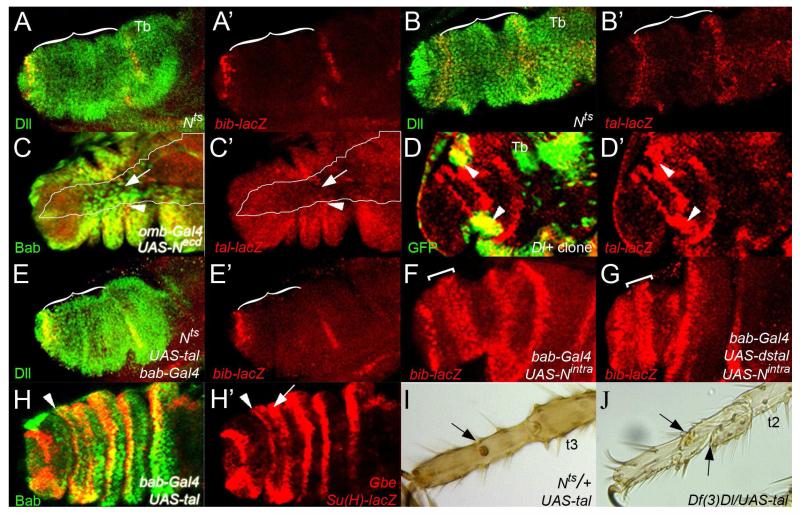
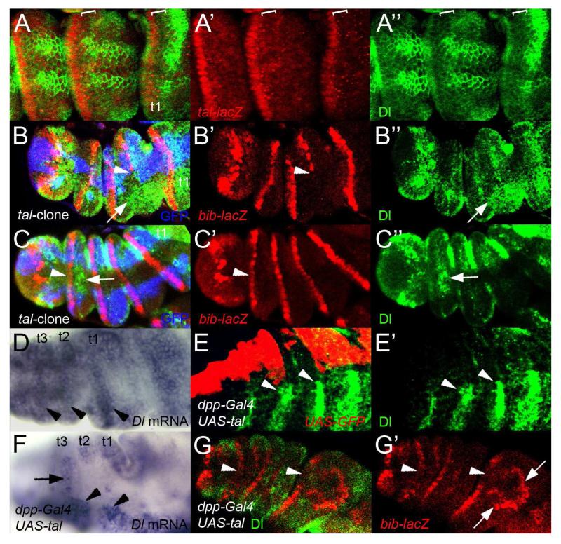
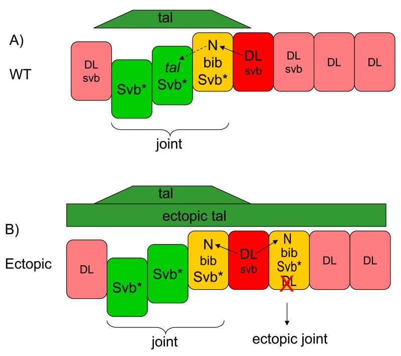
The formation of signalling boundaries is one of the strategies employed by the Notch (N) pathway to give rise to two distinct signalling populations of cells. Unravelling the mechanisms involved in the regulation of these signalling boundaries is essential to understanding the role of N during development and diseases. The function of N in the segmentation of the Drosophila leg provides a good system to pursue these mechanisms at the molecular level. Transcriptional and post-transcriptional regulation of the N ligands, Serrate (Ser) and Delta (Dl) generates a signalling boundary that allows the directional activation of N in the distalmost part of the segment, the presumptive joint. A negative feedback loop between odd-skipped-related genes and the N pathway maintains this signalling boundary throughout development in the true joints. However, the mechanisms controlling N signalling boundaries in the tarsal joints are unknown. Here we show that the non-canonical tarsal-less (tal) gene (also known as pri), which encodes for four small related peptides, is expressed in the N-activated region and required for joint development in the tarsi during pupal development. This function of tal is both temporally and functionally separate from the tal-mediated tarsal intercalation during mid-third instar that we reported previously. In the pupal function described here, N signalling activates tal expression and reciprocally Tal peptides feedback on N by repressing the transcription of Dl in the tarsal joints. This Tal-induced repression of Dl is mediated by the post-transcriptional activation of the Shavenbaby transcription factor, in a similar manner as it has been recently described in the embryo. Thus, a negative feedback loop involving Tal regulates the formation and maintenance of a Dl+/Dl- boundary in the tarsal segments highlighting an ancient mechanism for the regulation of N signalling based on the action of small cell signalling peptides.


Figures

Figure 1
Figure 1. Tal is required for joint development in the tarsal segments
A- Pupal leg (4h after puparium formation (APF)) showing stripes of tal mRNA expression in the tarsus (arrowheads). The leg is starting to evert; distal to the right. B- tal mRNA localisation in a pupal leg at 6h APF. tal is expressed in the distal part of the tarsal (t1-t4) segments (arrowheads) near the joint constrictions. C- Distal part of a tal-lacZ pupal leg (8h APF) showing strong tal expression in the distalmost part of the tarsal segments. D- D′′- Everted pupal leg (8h APF) showing tal mRNA (green, arrowhead) and bib-lacZ (red) expression patterns. bib-lacZ and tal mRNA are expressed adjacent to each other in the distalmost part of the tarsal segments. (D′) red channel showing bib-lacZ expression. (D′′) tal mRNA expression (arrowhead). E- Leg of a wild-type fly, showing the true segments (Coxa (Co), Trochanter (Tr), Femur (Fe), Tibia (Tb) and Pretasus (c)), and the tarsal segments (Ta). F- Distalmost part of a wild-type leg showing the tarsal segments (t1-t5) separated by naked joint tissue. Inset denotes the stereotypical ball and socket tarsal joint (arrowhead). Proximal to the top, distal to the bottom. G- Tarsal region of a bab-Gal4;UAS-dstal leg. The tarsal segments are misshapen, lacking joints (arrowheads). H- tal null clone induced between 96-120h AEL that runs along the ventral part of the tarsi marked with forked (blue). Note that every tarsal segment is still present. I- High magnification of H. In the tal null homozygous clone the joints do not form (arrowheads). However, some forked mutant bristles lacking tal can form part of remaining joint structures, revealing the non-autonomous nature of tal function (arrows). J- bab-Gal;UAS-tal tarsi showing ectopic joint structures (arrows) adjacent to the proper joint (arrowheads).
Figure 2
Figure 2. Tal regulates N target genes non-autonomously in the tarsal joint
A- A′- Expression of the joint marker bib-lacZ (red) in the tarsal region covered by Bab protein (green) in a pupal leg (6h APF) (A). bib-lacZ expression is limited to a single row of cells in the distal part of the tarsal segments (A′). B- B′- Distal part of a bib-lacZ pupal leg (5h APF) containing Minute+ GFP−, tal− null clones. GFP expression labels the tal+ tissue. bib-lacZ expression (red) is absent in the middle of a large tal mutant clone (brackets). Note that tal acts non-autonomously in bib-lacZ regulation (arrowhead) (B). Expression of bib-lacZ (B′). C- C′- A bib-lacZ (red) pupal leg (5h APF) expressing UAS-dstal in the tarsi using the bab-Gal4 driver (green) (C). Strong reduction of the bib-lacZ reporter is observed (arrows) (C′). D- D′- Ectopic joints in a bab-Gal4;UAS-tal pupal leg (6h APF) showing Bab (green) and bib-lacZ (red) patterns of expression (D). A duplicated row of bib-lacZ expressing cells is observed (arrowhead: endogenous; arrow: ectopic) (D′). E- E′- tal gain of function clones (green) induce ectopic expression (arrow) of the bib-lacZ reporter (red) in the distal part of the first tarsal segment (E). bib-lacZ expression (E′).
Figure 3
Figure 3. N signalling functions upstream and downstream of Tal in joint development
A- A′- Distal part of a Nts mutant pupal leg (4h APF) shifted to the restrictive Ta at late third instar, showing the unaffected expression of the Dll gene (green), that is not regulated by N, and bib-lacZ (red). No bib-lacZ expression is detected in the tarsus (brackets). (A′) red channel. B- B′- Nts mutant pupal leg (6h APF) treated as in A) showing tal-lacZ (red) and Dll (green) expression patterns (B). tal-lacZ expression is missing in the tarsus (brackets) (B′). C- C - Ectopic expression of a dominant negative N form (Necd) using omb-Gal4 driver in a pupal leg (6h APF) showing tal-lacZ (red) and Bab (green). Note that only Bab is expressed in the dorsal part of the disc (arrow) and tal-lacZ expression is repressed from this domain and only detected in the lateral parts (arrowhead) outside of omb-Gal4 domain (outline) (C). tal-lacZ expression (C′). D- D′- tal-lacZ (red) pupal leg (4h APF) containing Dl gain of function clones (green). Ectopic expression of tal-lacZ is observed in the Dl-expressing clones (arrowheads) (D). tal-lacZ expression (D′). E- E′- Overexpression of tal in a Nts mutant background pupal leg (4h APF) showing Dll (green) and bib-lacZ (red) expression patterns (E). bib-lacZ expression is lost in the tarsal segments (brackets) (E′). F- F′- Pupal leg (5h APF) expressing Nintra in the tarsal region. Nintra expands bib-lacZ expression in the tarsus (bracket). G- Ectopic co-expression of Nintra and UAS-dstal in the tarsi of a pupal leg (5h APF). bib-lacZ still appears expanded in the tarsus (bracket). H- H′- A 6h APF bab-Gal4;UAS-tal pupal leg showing the expression of an enhancer regulated directly by Nintra (Gbe+Su(H)-lacZ) (red) and Bab (green). Ectopic expression of Gbe+Su(H)-lacZ is observed in t4 in a row of cells (arrow) more proximal to the endogenous pattern (arrowhead); compare with Figs. 2D-E (H). Gbe+Su(H)-lacZ expression (H′). I- Distal part of a bab-Gal4;UAS-tal leg in a Nts heterozygous background. No ectopic joint structures are detected. Instead some tarsus display defective joints (arrow). J- Tarsi of a leg over-expressing tal in a heterozygous background for Dl (Df(3)DlBX12/+). As in I), no ectopic joint tissue is observed and some tarsal joints are defective, leading to tarsal segment fusions (arrows).
Figure 4
Figure 4. Tal represses Dl transcription in the N-responsive region to form a signalling border
A- A′′- Patterns of expression of Dl (green) and tal-lacZ (red) in the distal part of a pupal leg (6h APF) Note that Dl and tal-lacZ patterns of expression do not overlap (brackets) (A). tal-lacZ expression in the distalmost part of the segment (A′). Dl distribution showing a sharp boundary with non-Dl-expressing cells at the distal part of the segment (brackets) (A′′). B- B′′- 6h APF pupal leg showing a large Minute+ tal- mutant clone of around 10-15 cells wide (marked by lack of GFP, blue) and stained for bib-lacZ (red) and Dl expression (green) (B). bib-lacZ is lost in the larger area of the clone. Arrowhead denotes the edge of bib-lacZ expression. (B′) Dl distribution in this area does not form a boundary and it appears more distally (arrow) (B′′) C- C′′- Average Minute + tal− mutant clone in a 6h APF pupal leg labelled as in B). bib-lacZ expression is normal in the tal mutant clone (arrowhead) (C′). Dl is localized proximally forming a clear boundary (arrow; compare with B′′) (C′′). D- Dl mRNA distribution in a 5h APF pupal leg. Dl is highly expressed near the distal part of the segment (arrowheads), but is absent or at low concentration in the distalmost part. E- E′- Pupal leg (6h APF) over-expressing UAS-tal and UAS-GFP driven by dpp-Gal4, showing the GFP distribution in the dpp pattern (red) and Dl protein (green) (E). Dl protein (green) is only detected outside of the tal over-expressing domain (arrowheads) (E′). F- in situ hybridisation using a Dl riboprobe in a dpp-Gal4;UAS-tal pupal leg 5h APF. Dl transcription is repressed in tal over-expressing cells (arrow). Dl expression is detected outside the dpp pattern (arrowheads). G- G′- Ectopic expression of tal in a 5h APF pupal leg with dpp-Gal4 driver represses Dl (green) and bib-lacZ (red) in the dorsal part of the disc (arrowheads) but induces ectopic expression domains of bib-lacZ at the edges of the dpp-Gal4 domain (arrows) (G). Expression of bib-lacZ (G′).
Figure 5
Figure 5. Tal regulates N signalling through the Svb transcription factor
A- Distribution of svb mRNA in a 4h APF pupal leg. svb is detected in stripes in the tarsal segments. B- Tarsal joints of a svb107 mutant escaper displaying an incomplete joint (arrowhead). C- A svbR9 mutant clone marked with yellow in the tarsal segments (outlined in red). The cells lacking svb do not form autonomously the joint fold (arrowhead). D- Leg of a fly over-expressing UAS-svb in the tarsi is completely wild-type. E- Leg over-expressing both UAS-tal and UAS-svb using the bab-Gal4 driver. Apart from an abnormal joint in t1 (arrow) only attempts of joints can be observed in the rest of the tarsus (arrowhead). F- Distal part of a leg over-expressing an active Svb form (ovo-B) in the tarsi. The tarsal region is reduced and all joints are completely absent. G- G′′- Distribution of a GFP-tagged Svb protein (Svb-GFP) (red) in a tarsal segment of a pupal leg using the bab-Gal4 driver, which is expressed evenly throughout the tarsus. Expression of the bib-lacZ (green) indicates the proximal part of the joint region. (G). Svb-GFP is differentially distributed throughout the segment. In the joint region (thin brackets) Svb-GFP is strongly detected in entire nuclei (arrows) whereas in the proximal (non-joint) part of the segment (thick brackets), Svb-GFP is only detected in puncta (arrowheads) (G′). bib-lacZ expression (G′′). H- H′′- A 5h APF pupal leg showing svb-lacZ (red; arrowhead) and Dl (green; arrow) patterns of expression. Note that these adjacent expression domains are slightly overlapping (H). svb-lacZ reporter is expressed in the distal part of the tarsal segments (arrowheads) (H′). Dl protein distribution (arrow) (H′′). I- A bab-Gal4;UAS-tal;UAS-svb pupal leg (5h APF) showing a strong reduction of bib-lacZ expression in the tarsi (brackets). J- A pupal leg (5h APF) over-expressing ovo-B in the tarsi. The bib-lacZ pattern of expression is completely lost or very reduced (brackets). K- in situ hybridisation showing the Dl transcript pattern in a dpp-Gal4;UAS-ovoB pupal leg (4h APF). Dl expression is reduced in the dorsal part of the disc (arrow) but stripes of Dl are observed outside the dpp-Gal4 domain (arrowheads). L- L′′- A 5h APF pupal leg expressing ectopically ovo-B using the dpp-Gal4 driver (white outline) showing Dl protein distribution (green) and bib-lacZ expression (red). Arrow denotes the dorsal side of the disc where Dl is reduced and bib-lacZ expression is absent; the arrowhead marks the lateral side where bib-lacZ and Dl are present (L). Dl protein expression (L′). bib-lacZ expression (L′′).
Figure 6
Figure 6. Diagram depicting a model for the interactions between Tal and N during tarsal joint development
Schematic representation of the Tal-mediated mechanism controlling the formation of the N signalling boundary in the distal part of a tarsal segment in pupal legs (the orientation of the represented tarsal segments is as in other figure panels, distal to the left and proximal to the right). A negative feedback between N and Tal signalling regulates the formation and maintenance of N-signalling boundary. Joints (endogenous or ectopic) only arise if a) both tal and svb expression overlap (green) and b) this tal-svb overlap in turn abuts or overlaps high Dl expression (red; overlaps in yellow). These overlaps leads to the generation of a sharp Dl+/Dl− signalling boundary. A) By the end of third larval instar, Dl (red) and svb are expressed in slightly overlapping patterns in the distal part of the segment. At the onset of puparation, cells high levels of Dl and activate N signalling in the adjacent cells (black arrow). N signalling activates (directly or indirectly) tal gene expression in the distalmost part of the joint region (black dashed arrow). Non-autonomous Tal signalling (green) triggers the post-transcriptional activation the Svb transcription factor (Svb*) across the presumptive joint region (bracket). Subsequently, this Tal-mediated activation of Svb results in the direct or indirect transcriptional repression of Dl in the presumptive joint cells (yellow), generating a sharp Dl+/Dl− signalling boundary that leads to the activation of bib and other joint-promoting genes. Loss of tal or svb function results in the loss of this boundary, and hence, of joints. B) UAS-tal-mediated ectopic joints only arise in the region where endogenous Svb is present but out of reach of the endogenous Tal source, and yet overlapping or abutting the distal stripe of high Dl: these three conditions are only met in a narrow stripe proximal to the endogenous presumptive joint. Activation of Svb in this territory represses Dl and leads to the generation of a new Dl+/Dl− signalling boundary and the formation of an extra joint (arrows). Co-over-expression of UAS-svb plus UAS-tal, or expression of the Svb-activated form ovo-B throughout the segment eliminates Dl expression and precludes the formation of Dl+/Dl− signalling borders and joints.

Similar articles

Cited by

References

    1. Artavanis-Tsakonas S, Rand MD, Lake RJ. Notch signaling: cell fate control and signal integration in development. Science. 1999;284:770–6. - PubMed
    1. Bailey AM, Posakony JW. Suppressor of hairless directly activates transcription of enhancer of split complex genes in response to Notch receptor activity. Genes Dev. 1995;9:2609–22. - PubMed
    1. Becam I, Fiuza UM, Arias AM, Milan M. A role of receptor Notch in ligand cis-inhibition in Drosophila. Curr Biol. 2010;20:554–60. - PubMed
    1. Bishop SA, Klein T, Arias AM, Couso JP. Composite signalling from Serrate and Delta establishes leg segments in Drosophila through Notch. Development. 1999;126:2993–3003. - PubMed
    1. Blanpain C, Lowry WE, Pasolli HA, Fuchs E. Canonical notch signaling functions as a commitment switch in the epidermal lineage. Genes Dev. 2006;20:3022–35. - PMC - PubMed

Publication types

MeSH terms