. 2011 May 27;332(6033):1089-92.
doi: 10.1126/science.1205358. Epub 2011 Apr 28.

Structural basis for methyl transfer by a radical SAM enzyme

Affiliations

Structural basis for methyl transfer by a radical SAM enzyme

Amie K Boal et al. Science. .

Abstract

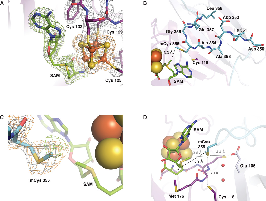
The radical S-adenosyl-L-methionine (SAM) enzymes RlmN and Cfr methylate 23S ribosomal RNA, modifying the C2 or C8 position of adenosine 2503. The methyl groups are installed by a two-step sequence involving initial methylation of a conserved Cys residue (RlmN Cys(355)) by SAM. Methyl transfer to the substrate requires reductive cleavage of a second equivalent of SAM. Crystal structures of RlmN and RlmN with SAM show that a single molecule of SAM coordinates the [4Fe-4S] cluster. Residue Cys(355) is S-methylated and located proximal to the SAM methyl group, suggesting the SAM that is involved in the initial methyl transfer binds at the same site. Thus, RlmN accomplishes its complex reaction with structural economy, harnessing the two most important reactivities of SAM within a single site.


Figures

Fig. 1
Fig. 1
The structure of E. coli RlmN with S-adenosyl-L-methionine (SAM) (A). The α66 partial barrel core is shown in dark purple, a three strand β extension to the core (β’1–3 extension) is shown in light purple, an N-terminal helical accessory domain is shown in green, and a C-terminal extension (β7 extension) culminating in an α-helix is shown in blue. The [4Fe-4S] cluster cofactor is represented as a space-filling model and SAM is shown in stick format colored by atom type. Cysteine (Cys) ligands to the [4Fe-4S] cluster and mechanistically important residues Cys 118 and methylcysteine (mCys) 355 are shown as purple and blue sticks, respectively, and colored by atom type. (B) A top-down view comparing the RlmN active site in the absence (right) and presence (left) of SAM. An unmodeled region (residues 351–360) in the RlmN structure obtained without SAM is represented as a dashed line.
Fig. 2
Fig. 2
Selected views of the RlmN active site. (A) 2Fo-Fc electron density map (gray mesh, 1.5σ) for residues 125–132 (containing the CX3CX2C motif), the [4Fe-4S] cluster, and the SAM cosubstrate in the RlmN+SAM structure. An omit map contoured at 3.0σ for SAM (green mesh) and the [4Fe-4S] cluster (orange mesh) is superimposed. (B) A top-down view of the highly conserved 351–360 linker region as modeled in the RlmN+SAM structure. The linker is represented in stick format colored by atom type. A loop (residues 310–320) located in front of the SAM cofactor is omitted for clarity. (C) 2Fo-Fc electron density map (gray mesh, 1.0σ) for residue 355 in the RlmN+SAM structure. Omit maps for the S-methyl group of residue 355 (green mesh, 2.2σ) and the entire residue (orange mesh, 1.9σ) are superimposed. (D) A side view of the active site. The conserved MGMGE sequence motif in the α66 core is shown in stick format and colored by atom type. Selected conserved residues and ordered water molecules are shown as sticks and spheres, respectively. The 5’ position of the SAM cosubstrate is denoted by an asterisk. Distances relevant to the methyl transfer reaction are drawn as gray lines and those relevant to mCys 355 activation and disulfide formation are shown as black lines.
Fig. 3
Fig. 3
Electrostatic surface potential (A, B) and Cfr sequence conservation (C, D) maps of the RlmN structure. A Electrostatic surface calculated with APBS (47) contoured at -15 kBT (red, negative) and 15 kBT (blue, positive). (B) View rotated 90° about the vertical axis and 90° about the horizontal axis from view (A). (C) Map of sequence conservation between E. coli RlmN and S. aureus Cfr using a pairwise alignment. Strictly conserved residues are shown in orange, neutral substitutions are shown in tan, and variable regions are shown in white. Residues 344–350 from the β7 extension (which includes conserved residues Arg 344, Gly 348, and Asp 350) are omitted to afford a better view of the active site cavity. (D) View rotated 90° about the vertical axis and 90° about the horizontal axis from view (C).

References

    1. Markham GD. Encyclopedia of Life Sciences. John Wiley & Sons, Inc; 2010.
    1. Woodard RW, Tsai MD, Floss HG, Crooks PA, Coward JK. J. Biol. Chem. 1980;255:9124. - PubMed
    1. Hegazi MF, Borchardt RT, Schowen RL. J. Am. Chem. Soc. 1979;101:4359.
    1. Frey PA, Hegeman AD. Enzymatic Reaction Mechanisms. Oxford University Press; New York: 2007.
    1. Iwig DF, Grippe AT, McIntyre TA, Booker SJ. Biochemistry. 2004;43:13510. - PubMed

Publication types

MeSH terms