. 2011 Jul;301(1):G110-8.
doi: 10.1152/ajpgi.00412.2010. Epub 2011 Apr 28.

Hepatic stellate cells require a stiff environment for myofibroblastic differentiation

Affiliations

Hepatic stellate cells require a stiff environment for myofibroblastic differentiation

Abby L Olsen et al. Am J Physiol Gastrointest Liver Physiol. 2011 Jul.

Abstract

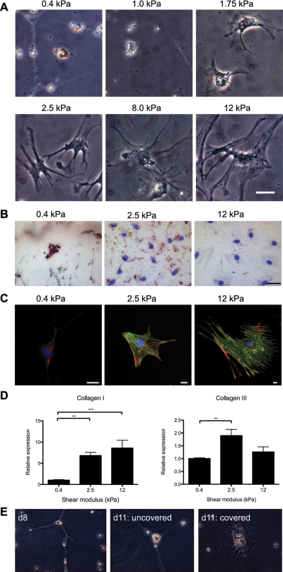
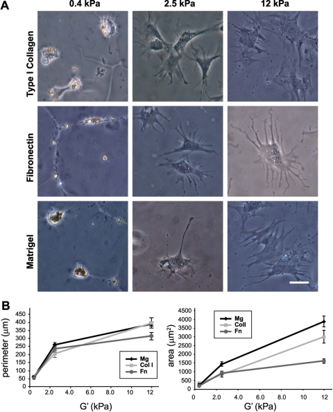
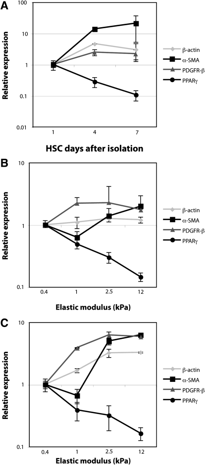
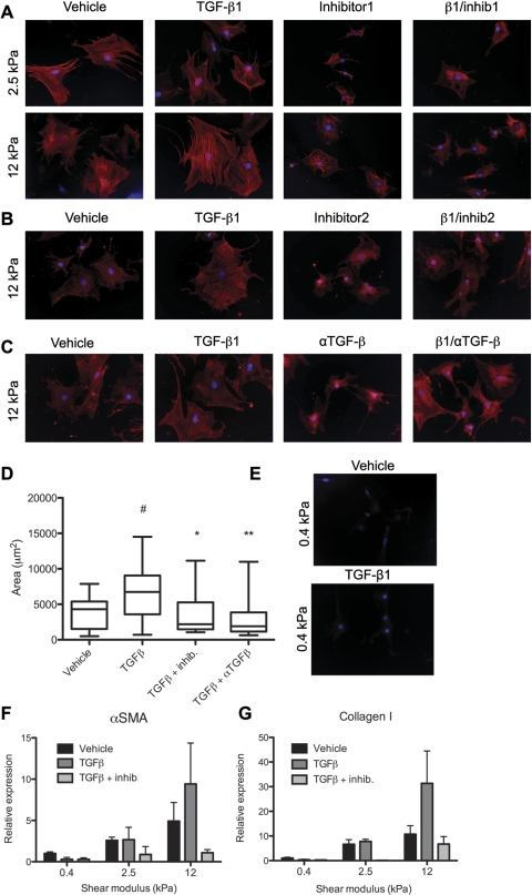
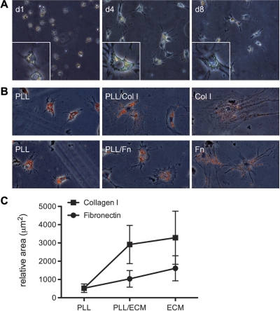
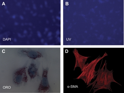
The myofibroblastic differentiation of hepatic stellate cells (HSC) is a critical event in liver fibrosis and is part of the final common pathway to cirrhosis in chronic liver disease from all causes. The molecular mechanisms driving HSC differentiation are not fully understood. Because macroscopic tissue stiffening is a feature of fibrotic disease, we hypothesized that mechanical properties of the underlying matrix are a principal determinant of HSC activation. Primary rat HSC were cultured on inert polyacrylamide supports of variable but precisely defined shear modulus (stiffness) coated with different extracellular matrix proteins or poly-L-lysine. HSC differentiation was determined by cell morphology, immunofluorescence staining, and gene expression. HSC became progressively myofibroblastic as substrate stiffness increased on all coating matrices, including Matrigel. The degree rather than speed of HSC activation correlated with substrate stiffness, with cells cultured on supports of intermediate stiffness adopting stable intermediate phenotypes. Quiescent cells on soft supports were able to undergo myofibroblastic differentiation with exposure to stiff supports. Stiffness-dependent differentiation required adhesion to matrix proteins and the generation of mechanical tension. Transforming growth factor-β treatment enhanced differentiation on stiff supports, but was not required. HSC differentiate to myofibroblasts in vitro primarily as a function of the physical rather than the chemical properties of the substrate. HSC require a mechanically stiff substrate, with adhesion to matrix proteins and the generation of mechanical tension, to differentiate. These findings suggest that alterations in liver stiffness are a key factor driving the progression of fibrosis.


Figures

Fig. 1.
Fig. 1.
Hepatic stellate cells (HSC) demonstrate increased spreading and α-smooth muscle actin (α-SMA) expression on stiffer substrates. A: freshly isolated rat HSC were cultured for 7 days on type I collagen-coated polyacrylamide gels of varying shear modulus (G′), ranging from 0.4 to 12 kPa. By light microscopy, HSC appear morphologically quiescent on soft supports (0.4–1.0 kPa). These cells also demonstrated UV autofluorescence (data not shown). HSC plated on stiff (8–12 kPa) polyacrylamide supports displayed an activated, myofibroblastic-like phenotype. HSC cultured on intermediate supports (1.75–2.5 kPa) showed intermediate phenotypes. These results are representative of 3 experiments. Bar, 50 μm. B: HSC were cultured as in A and were stained with Oil Red O (ORO) and counterstained with 4,6-diamidino-2-phenylindole (DAPI; blue). Lipid droplets decrease on stiffer gels. Representative cells are from 2 different experiments Bar, 50 μM. C: HSC were cultured as in A and were immunostained with antibodies against the rat stellate cell marker desmin (red) and α-SMA (green); nuclei were stained with DAPI (blue). Desmin expression decreases in cells on stiffer gels, whereas α-SMA expression and cell size increase. α-SMA is organized in stress fibers in cells on 12-kPa gels. Photos are representative of 3 experiments (bars, 10 μm, note different magnifications). D: HSC were cultured as in A, and quantitative RT-PCR (qRT-PCR) for collagen I and collagen III was performed. Results are normalized to expression of 18s rRNA and are averaged from 3–4 independent experiments, each done in triplicate. Values are means ± SE. **P < 0.01. ***P < 0.005. E: primary HSC were cultured for 8 days (d8) on type I collagen-coated polyacrylamide gels of 0.4-kPa stiffness, maintaining a quiescent phenotype (left). Cells cultured under the same conditions for an additional 3 days remained quiescent in appearance (middle), whereas cells on a region of the gel covered with a glass coverslip for the additional 3 days began to spread and lose lipid droplets (right). Representative cells are shown from 2 independent experiments; ×40 magnification.
Fig. 2.
Fig. 2.
HSC undergo myofibroblastic differentiation on stiff substrates, independent of the matrix coating. A: HSC were cultured for 7 days on polyacrylamide gels of varying stiffness coated with type I collagen, plasma fibronectin, or Matrigel. Regardless of the extracellular matrix (ECM) coating, HSC adopted a myofibroblastic phenotype on rigid (12 kPa) supports, and they remained quiescent on soft (0.4 kPa) supports. Results are representative of at least 3 experiments. Bar, 50 μm. B: cells on the different matrices and supports were traced using Image J software for quantification of perimeter (left) and area (right). Values are means ± SD. Mg, Matrigel; Col I, type I collagen; Fn, plasma fibronectin. A minimum of 40 cells were analyzed for each condition.
Fig. 3.
Fig. 3.
Changes in gene expression on increasingly stiff substrates parallel those occurring on plastic over time. A: primary HSC were cultured on tissue culture plastic and harvested on day 1 (quiescent phenotype), day 4 (intermediate), and day 7 (myofibroblastic). mRNA expression of four genes [β-actin, α-SMA, PDGF receptor-β (PDGF-Rβ), and peroxisome proliferator-activated receptor-γ (PPAR-γ)] was assayed by qRT-PCR and normalized to the 18S rRNA internal control. Changes in gene expression are shown relative to expression levels at day 1. This experiment was performed in duplicate, with 2 independent isolates of HSC (n = 4 replicates total). B and C: primary HSC were cultured for 7 days on polyacrylamide supports of variable stiffness, coated with either 0.1 mg/ml type I collagen (B) or 0.2 mg/ml plasma fibronectin (C). mRNA expression of the same panel of genes was determined by qRT-PCR, with levels at each point expressed relative to those for soft supports (0.4 kPa). With increasing stiffness of polyacrylamide supports, changes in HSC gene expression paralleled those observed with progressive HSC activation. Each experiment (B and C) was performed in duplicate, with 3 independent isolates of cells (n = 6 total). Values are means ± SD.
Fig. 4.
Fig. 4.
HSC on stiff substrates express α-SMA, even in the absence of transforming growth factor (TGF)-β. Primary rat HSC were cultured on type I collagen-coated polyacrylamide substrates of 2.5 or 12 kPa. A: beginning on the day after isolation, after cells adhered to the gels, they were treated with either vehicle, 100 pM TGF-β1, 5 μM TGF-β kinase inhibitor (NPC-34016; inhibitor 1), or a combination of TGF-β and the kinase inhibitor. After 7 days, cells were fixed and stained with antibody against α-SMA (red). Nuclei are stained with DAPI (blue). ×20 Magnification. B and C: cells were treated as in A, but with a different kinase inhibitor (616451; inhibitor 2) or a pan-TGF-β blocking antibody (αTGF-β). Cells were stained as in A. Results are representative of 2 independent experiments. Controls were treated with vehicle and mouse serum IgG. D: cell area was calculated using National Institutes of Health Image J. Results are representative of 2 independent experiments. Values are means ± SE. #Significantly different from vehicle, P < 0.05. *Significantly different from TGF-β treatment, P < 0.05. **Significantly different from TGF-β treatment, P < 0.01. E: primary rat HSC were cultured on type I collagen-coated polyacrylamide substrates of 0.4-kPa stiffness. Beginning on the day after isolation, after cells adhered to the gels, they were treated with either vehicle or 100 pM TGF-β1. After 7 days, cells were fixed and stained with antibody against α-SMA (red). Nuclei are stained with DAPI (blue). ×20 Magnification. F and G: cells were treated as in B, then analyzed by real-time PCR for expression of α-SMA (F) and type I collagen (G). Values are means ± SE.
Fig. 5.
Fig. 5.
HSC require matrix protein interactions to undergo myofibroblastic differentiation on stiff supports. A: primary rat HSC were cultured on 12-kPa polyacrylamide supports coated with 0.1 mg/ml poly-l-lysine (PLL) for up to 8 days. Even at day 8, the cells are minimally spread and retain vitamin A droplets (bright field). Photos are representative of cells in 4 experiments. ×10 Magnification. B: primary rat HSC were cultured for 7 days on 0.1 mg/ml PLL alone, on a mixture of 0.05 mg/ml PLL and 0.05 mg/ml type I collagen or plasma fibronectin, or on 0.1 mg/ml collagen or plasma fibronectin alone. ORO staining demonstrates retention of lipid droplets in cells on PLL, but increasing spreading and loss of lipid droplets with increasing amounts of matrix proteins. ×20 Magnification. C: quantification of the cell area in B, where ECM represents either collagen or fibronectin. Cells (15–32 per data point) were traced with Image J to determine area, with data shown as means ± SD. Two-way ANOVA demonstrated statistical significance for PLL and PLL/ECM points compared with PLL alone, with P < 0.0001.
Fig. 6.
Fig. 6.
HSC require the generation of mechanical tension to undergo myofibroblastic differentiation on stiff supports. Primary rat HSC were cultured on Teflon supports for 7 days. A: nuclei were stained with DAPI (blue). B: vitamin A-containing lipid droplets were visualized by UV autofluorescence. ×20 Magnification. C and D: cells treated as above were gently removed from the Teflon by rinsing and were replated on glass. C: after 6 h, cells stained with ORO demonstrated persistent lipid droplets. ×60 Magnification. D: cells cultured for 5 days on glass (after 7 days on Teflon), then stained with antibodies against α-SMA (red), demonstrated α-SMA in stress fibers. ×20 Magnification. ORO and immunostains are representative of 3 independent experiments.

References

    1. Arora PD, Narani N, McCulloch CA. The compliance of collagen gels regulates transforming growth factor-beta induction of alpha-smooth muscle actin in fibroblasts. Am J Pathol 154: 871–882, 1999 - PMC - PubMed
    1. Basu A, Wen Q, Lubensky T, Janmey P, Yodh A. Non-affine displacements in flexible polymer gels. Macromol. 44: 1671–1679, 2011
    1. Bershadsky A, Kozlov M, Geiger B. Adhesion-mediated mechanosensitivity: a time to experiment, and a time to theorize. Curr Opin Cell Biol 18: 472–481, 2006 - PubMed
    1. Bissell DM, Wang SS, Jarnagin WR, Roll FJ. Cell-specific expression of transforming growth factor-beta in rat liver. Evidence for autocrine regulation of hepatocyte proliferation. J Clin Invest 96: 447–455, 1995 - PMC - PubMed
    1. Bridle KR, Crawford DH, Ramm GA. Identification and characterization of the hepatic stellate cell transferrin receptor. Am J Pathol 162: 1661–1667, 2003 - PMC - PubMed

Publication types

MeSH terms