

Peripheral giant cell granuloma: a review of 123 cases
- PMID: 21528029
- PMCID: PMC3075451
Peripheral giant cell granuloma: a review of 123 cases
Abstract
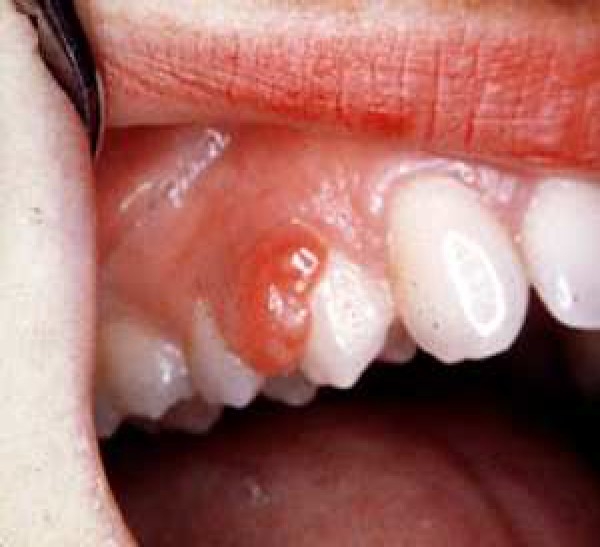
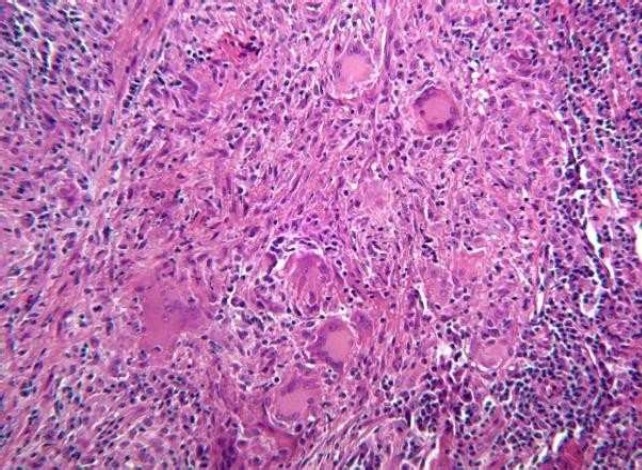
Background: Peripheral giant cell granuloma is one of the reactive hyperplastic lesions of the oral cavity, which originates from the periosteum or periodontal membrane following local irritation or chronic trauma. The purpose of this study was to present the clinical characteristics of peripheral giant cell granuloma in a group of Iranian population.
Methods: A series of 123 consecutive confirmed cases of peripheral giant cell granuloma after biopsy were evaluated. Age, sex, anatomic location, consistency, etiologic factor, pain and bleeding history, color, surface texture, and pedicle situation were recorded and were analyzed by chi-square test and values were considered to be significant if P < 0.05.
Results: Age ranged from 6 to 75 years (mean 33 years). Women affected more than men (M/F 1:1.1). Peripheral giant cell granuloma was seen in the mandible more than in the maxilla and in the anterior region more than in the posterior region. In most cases, lesions were pink, pedunculated and had non-ulcerated surface. In less than half of the cases, there was no history of bleeding and also pain was rarely reported. Calculus was the most common etiologic factor.
Conclusion: The results confirmed that the clinical features of peripheral giant cell granuloma in a group of Iranian population are almost similar to those reported by other investigators.
Keywords: Giant cell epulis; giant cell granuloma; trauma.
Figures
References
-
- Wood NK, Goaz PW. 5th ed. St.Louis: Mosby; 1997. Differential Diagnosis of Oral and Maxillofacial Lesions; pp. 141–2.
-
- Carvalho YR, Loyola AM, Gomez RS, Araujo VC. Peripheral giant cell granuloma. An immunohisto-chemical and ultrastructural study. Oral Dis. 1995;1(1):20–5. - PubMed
-
- Regezi JA SJJR. 5th ed. St.Louis: WB. Saunders; 2007. Oral Pathology: Clinical Pathologic Correlations; pp. 112–3.
-
- Chaparro-Avendano AV, Berini-Aytes L, Gay-Escoda C. Peripheral giant cell granuloma. A report of five cases and review of the literature. Med Oral Patol Oral Cir Bucal. 2005;10(1):53–7. - PubMed
-
- Motamedi MH, Eshghyar N, Jafari SM, Lassemi E, Navi F, Abbas FM, et al. Peripheral and central giant cell granulomas of the jaws: a demographic study. Oral Surg Oral Med Oral Pathol Oral Radiol Endod. 2007;103(3):e39–e43. - PubMed
LinkOut - more resources
Full Text Sources