

Intraoral epithelioid hemangioendothelioma: an intermediate vascular tumor- a case report
- PMID: 21528039
- PMCID: PMC3075463
Intraoral epithelioid hemangioendothelioma: an intermediate vascular tumor- a case report
Abstract
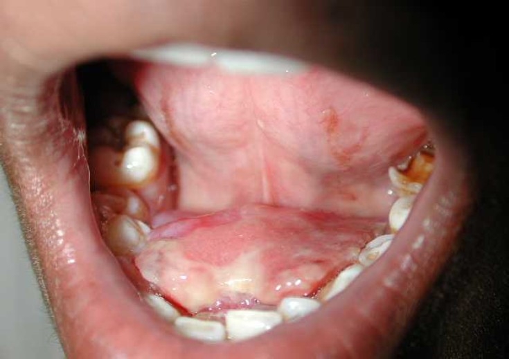
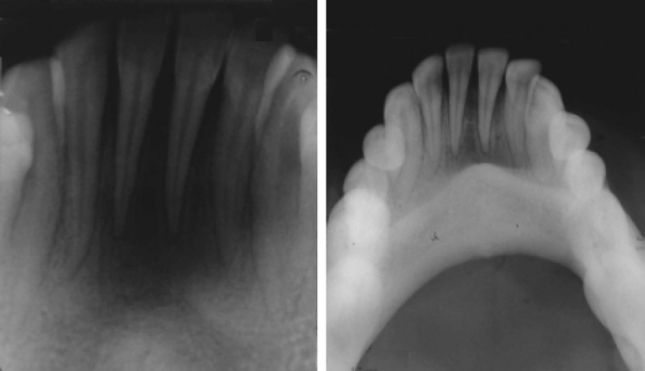
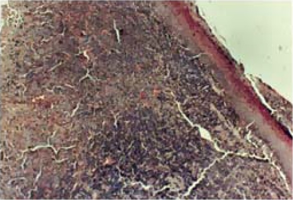
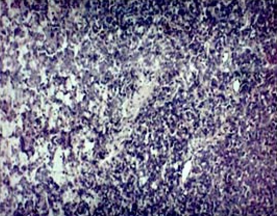
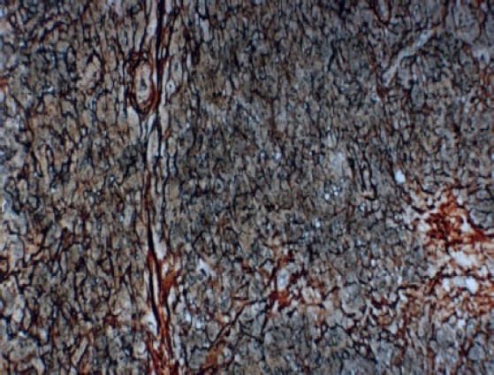
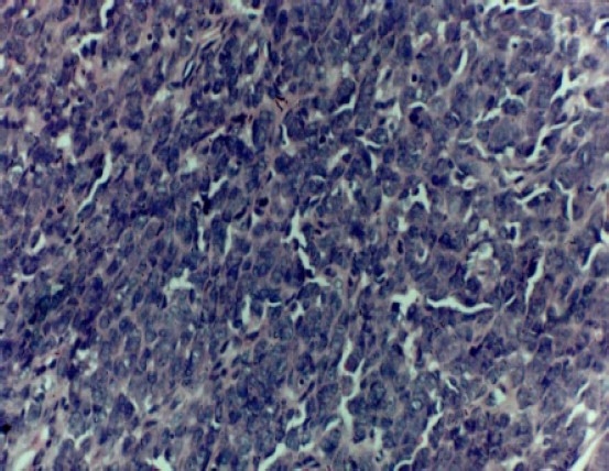
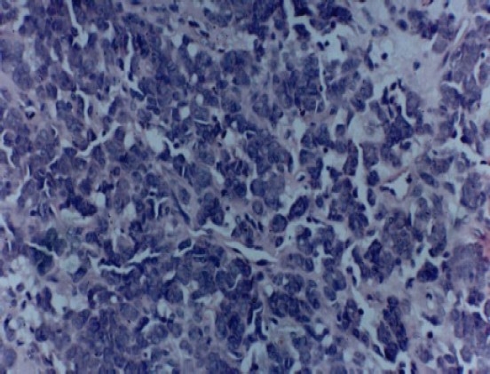
Vascular neoplasms, other than benign are characterized as intermediate or malignant. They are often enshrouded in controversy, because the same neoplasm could show variability in biologic behavior that may not be correlated with microscopic features. The intermediate grade vascular neoplasm is named as epithelioid hemangioendothelioma (EHE). Epithelioid hemangioendothelioma of the oral cavity has been infrequently reported. To the best of our knowledge, the review of the English literature revealed a total of 30 cases of intraoral EHE reported till today. We report such a rare case in a 20 year old male, presented with a growth in lower anterior lingual gingiva since five months before the diagnosis with a history of similar swelling, twice in the same area. The differential diagnosis and brief review of literature is also discussed in the current article.
Keywords: Epithelioid; Hemangioendothelioma; Hemangiosarcoma.
Figures
References
-
- Weiss SW, Enzinger FM. Epithelioid hemangioendothelioma: a vascular tumor often mistaken for a carcinoma. Cancer. 1982;50(5):970–81. - PubMed
-
- Enzinger FM, Weiss SW. 3rd ed. Philadelphia: Mosby; 1995. Soft tissue tumors; pp. 891–900.
-
- Wesley RK, Mintz SM, Wertheimer FW. Primary malignant hemangioendothelioma of the gingival. Report of a case and review of the literature. Oral Surg Oral Med Oral Pathol. 1975;39(1):103–12. - PubMed
-
- Marrogi AJ, Boyd D, el Mofty S, Waldron C. Epithelioid hemangioendothelioma of the oral cavity: report of two cases and review of literature. J Oral Maxillofac Surg. 1991;49(6):633–8. - PubMed
-
- Orsini G, Fioroni M, Rubini C, Piattelli A. Epithelioid hemangioendothelioma of the oral cavity: report of case. J Oral Maxillofac Surg. 2001;59(3):334–7. - PubMed
Publication types
LinkOut - more resources
Full Text Sources