

Cytochrome P450s in the regulation of cellular retinoic acid metabolism
- PMID: 21529158
- PMCID: PMC3789243
- DOI: 10.1146/annurev-nutr-072610-145127
Cytochrome P450s in the regulation of cellular retinoic acid metabolism
Abstract
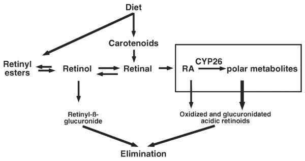
The active metabolite of vitamin A, retinoic acid (RA), is a powerful regulator of gene transcription. RA is also a therapeutic drug. The oxidative metabolism of RA by certain members of the cytochrome P450 (CYP) superfamily helps to maintain tissue RA concentrations within appropriate bounds. The CYP26 family--CYP26A1, CYP26B1, and CYP26C1--is distinguished by being both regulated by and active toward all-trans-RA (at-RA) while being expressed in different tissue-specific patterns. The CYP26A1 gene is regulated by multiple RA response elements. CYP26A1 is essential for embryonic development, whereas CYP26B1 is essential for postnatal survival as well as germ cell development. Enzyme kinetic studies have demonstrated that several CYP proteins are capable of metabolizing at-RA; however, it is likely that CYP26A1 plays a major role in RA clearance. Thus, pharmacological approaches to limiting the activity of CYP26 enzymes may extend the half-life of RA and could be useful clinically in the future.
Figures


References
-
- Abu-Abed S, MacLean G, Fraulob V, Chambon P, Petkovich M, Dollé P. Differential expression of the retinoic acid-metabolizing enzymes CYP26A1 and CYP26B1 during murine organogenesis. Mech. Dev. 2002;110:173–77. - PubMed
-
- Andreola F, Hayhurst GP, Luo G, Ferguson SS, Gonzalez FJ, et al. Mouse liver CYP2C39 is a novel retinoic acid 4-hydroxylase. Its down-regulation offers a molecular basis for liver retinoid accumulation and fibrosis in aryl hydrocarbon receptor-null mice. J. Biol. Chem. 2004;279:3434–38. - PubMed
-
- Ashla AA, Hoshikawa Y, Tsuchiya H, Hashiguchi K, Enjoji M, et al. Genetic analysis of expression profile involved in retinoid metabolism in non-alcoholic fatty liver disease. Hepatol. Res. 2010;40:594–604. - PubMed
-
- Balmer JE, Blomhoff R. Gene expression regulation by retinoic acid. J. Lipid Res. 2002;43:1773–808. - PubMed
Publication types
MeSH terms
Substances
Grants and funding
LinkOut - more resources
Full Text Sources
Other Literature Sources
Molecular Biology Databases
