Review
. 2011 Apr 29;145(3):341-55.
doi: 10.1016/j.cell.2011.04.005.

Macrophages in the pathogenesis of atherosclerosis

Affiliations
Review

Macrophages in the pathogenesis of atherosclerosis

Kathryn J Moore et al. Cell. .

Abstract

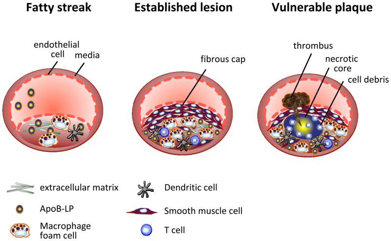
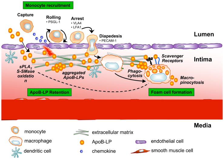
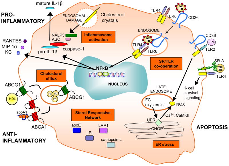
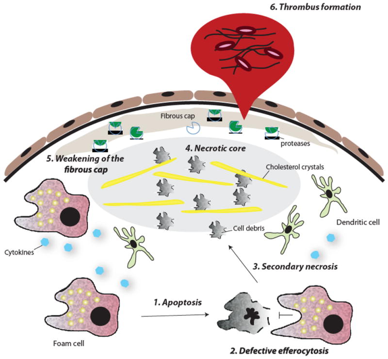
In atherosclerosis, the accumulation of apolipoprotein B-lipoproteins in the matrix beneath the endothelial cell layer of blood vessels leads to the recruitment of monocytes, the cells of the immune system that give rise to macrophages and dendritic cells. Macrophages derived from these recruited monocytes participate in a maladaptive, nonresolving inflammatory response that expands the subendothelial layer due to the accumulation of cells, lipid, and matrix. Some lesions subsequently form a necrotic core, triggering acute thrombotic vascular disease, including myocardial infarction, stroke, and sudden cardiac death. This Review discusses the central roles of macrophages in each of these stages of disease pathogenesis.


Figures

Figure 1
Figure 1. Progression of an atherosclerotic lesion
Early fatty streak lesions are characterized by the accumulation of apoB-LPs in the subendothelial space which incites the recruitment of dendritic cells and macrophages. As the atherosclerotic lesion progresses, smooth muscle and T cells also infiltrate the intima, and apoB-LP retention is amplified. Vulnerable plaques are characterized by the accumulation of apoptotic cells and defective efferocytosis resulting in the lipid filled necrotic core. A thinning fibrous cap decreases lesion stability making these atherosclerotic plaques susceptible to rupture and the formation of a thrombus.
Figure 2
Figure 2. Retention of apoB-LPs incites monocyte recruitment in early atherosclerotic lesions
ApoB-LPs enter the intima, bind to proteoglycans, and undergo various modifications, including oxidation and hydrolysis by sPLA2 and S-SMase. These modifications incite an inflammatory response characterized by chemokine secretion and altered expression of adhesion molecules by the overlying endothelial cells; cause lipoprotein aggregation; and further promote lipoprotein retention. The inflammatory signals lead to monocyte recruitment into the intima, where they differentiate into macrophages and internalize native and modified lipoproteins resulting in foam cell formation. Moreover, foam cells can contribute further to, and thus amplify, lipoprotein modifications and retention (dotted arrow).
Figure 3
Figure 3. Signaling pathways activated in the lesional macrophage
Various pro- and anti-inflammatory forces act on the macrophage in atherosclerotic lesions leading to activation of downstream cascades, such as the inflammasome, scavenger receptor (SR)/Toll-like receptor (TLR) cooperative signaling, ER stress, expression of the sterol responsive network, and efflux of cholesterol via ABCA1 and ABCG1 transporters.
Figure 4
Figure 4. Forces in the advanced lesion that destabilize the atherosclerotic plaque
(1) Macrophage foam cells undergo apoptosis as a result of prolonged ER stress and other stimuli; (2) these apoptotic cells are not effectively cleared by advanced lesional macrophages (defective efferocytosis); (3) accumulation of apoptotic cells induces secondary necrosis and (4) contributes to the formation of a necrotic core. (5) Smooth muscle cell death and protease degradation of extracellular matrix weakens the fibrous cap, making it susceptible to rupture. (6) Exposure of the thrombogenic material of the lesion causes platelet aggregation and thrombus formation.

References

    1. Abi-Younes S, Sauty A, Mach F, Sukhova GK, Libby P, Luster AD. The stromal cell-derived factor-1 chemokine is a potent platelet agonist highly expressed in atherosclerotic plaques. Circ Res. 2000;86:131–138. - PubMed
    1. Aouadi M, Tesz GJ, Nicoloro SM, Wang M, Chouinard M, Soto E, Ostroff GR, Czech MP. Orally delivered siRNA targeting macrophage Map4k4 suppresses systemic inflammation. Nature. 2009;458:1180–1184. - PMC - PubMed
    1. Arai S, Shelton JM, Chen M, Bradley MN, Castrillo A, Bookout AL, Mak PA, Edwards PA, Mangelsdorf DJ, Tontonoz P, Miyazaki T. A role for the apoptosis inhibitory factor AIM/Spα/Api6 in atherosclerosis development. Cell Metabolism. 2005;1:201–213. - PubMed
    1. Arnold L, Henry A, Poron F, Baba-Amer Y, Van RN, Plonquet A, Gherardi RK, Chazaud B. Inflammatory monocytes recruited after skeletal muscle injury switch into antiinflammatory macrophages to support myogenesis. J Exp Med. 2007;204:1057–1069. - PMC - PubMed
    1. Babaev VR, Chew JD, Ding L, Davis S, Breyer MD, Breyer RM, Oates JA, Fazio S, Linton MF. Macrophage EP4 deficiency increases apoptosis and suppresses early atherosclerosis. Cell Metab. 2008;8:492–501. - PMC - PubMed

Publication types