. 2011 May 13;88(5):536-47.
doi: 10.1016/j.ajhg.2011.04.003. Epub 2011 Apr 28.

Human mutations in NDE1 cause extreme microcephaly with lissencephaly [corrected]

Affiliations

Human mutations in NDE1 cause extreme microcephaly with lissencephaly [corrected]

Fowzan S Alkuraya et al. Am J Hum Genet. .

Erratum in

  • Am J Hum Genet. 2011 May 13;88(5):677

Abstract

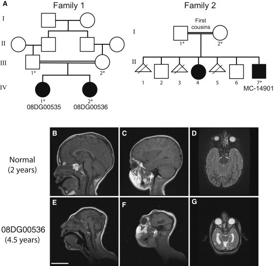
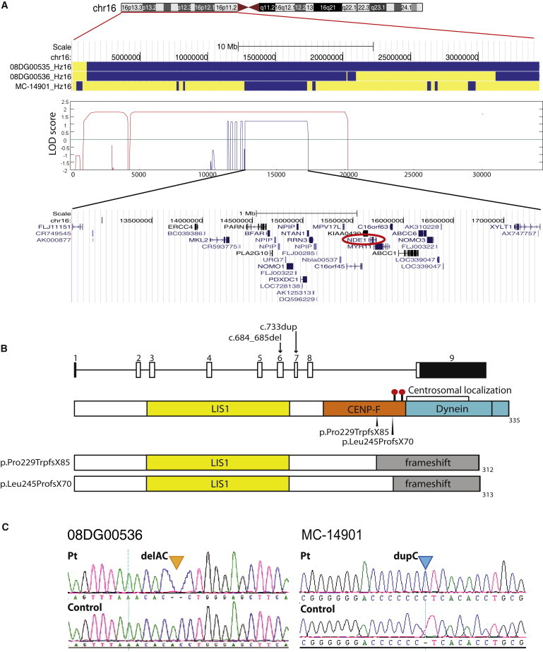
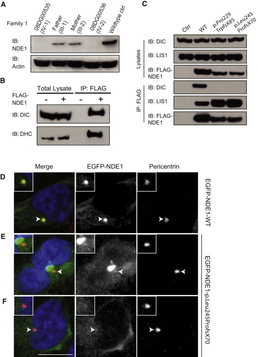
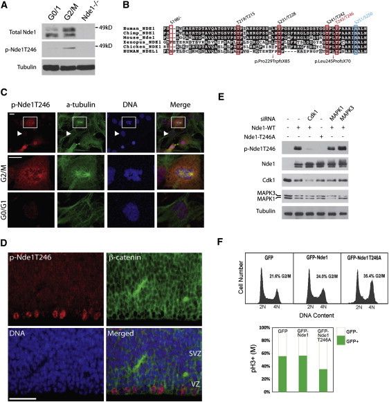
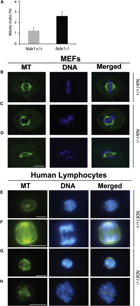
Genes disrupted in human microcephaly (meaning "small brain") define key regulators of neural progenitor proliferation and cell-fate specification. In comparison, genes mutated in human lissencephaly (lissos means smooth and cephalos means brain) highlight critical regulators of neuronal migration. Here, we report two families with extreme microcephaly and grossly simplified cortical gyral structure, a condition referred to as microlissencephaly, and show that they carry homozygous frameshift mutations in NDE1, which encodes a multidomain protein that localizes to the centrosome and mitotic spindle poles. Both human mutations in NDE1 truncate the C-terminal NDE1domains, which are essential for interactions with cytoplasmic dynein and thus for regulation of cytoskeletal dynamics in mitosis and for cell-cycle-dependent phosphorylation of NDE1 by Cdk1. We show that the patient NDE1 proteins are unstable, cannot bind cytoplasmic dynein, and do not localize properly to the centrosome. Additionally, we show that CDK1 phosphorylation at T246, which is within the C-terminal region disrupted by the mutations, is required for cell-cycle progression from the G2 to the M phase. The role of NDE1 in cell-cycle progression probably contributes to the profound neuronal proliferation defects evident in Nde1-null mice and patients with NDE1 mutations, demonstrating the essential role of NDE1 in human cerebral cortical neurogenesis.


Figures

Figure 1
Figure 1
Pedigrees and Radiographic Features of the Two Consanguineous Families with Microlissencephaly (A) Both families are from Saudi Arabia. Parents of family 1 are first cousins and have two affected female children (08DG00535, IV-1 and 08DG00536, IV-2). Whole-blood DNA from both parents and both affected children was obtained and analyzed (indicated by an asterisk). Parents of family 2 are first cousins who had seven reported pregnancies, producing one affected male (MC-14901, II-7), one affected female (II-4) (not available for analysis), two unaffected males, and three pregnancies that resulted in fetal demise. Whole-blood DNA from both parents and the affected male (MC-14901, II-7) was obtained and analyzed (indicated by an asterisk). (B–G) Representative MRI images of 08DG00536 (IV-2) from family 1 at 4.5 years of age, demonstrating the drastic reduction in brain size (E–G), agenesis of the corpus callosum, and abnormal gyral pattern compared to that seen in a normal 2-year-old child (B-D). Sagittal T1 (B, C, E, and F) and axial T2 (D and G) sections are shown. The scale bar represents 5 cm. Additional images are available as Movies S1–S6.
Figure 2
Figure 2
Two NDE1 Mutations Identified within an Overlapping Region of Homozygosity in Both Pedigrees on Chromosome 16 (A) Homozygosity analysis of both pedigrees identified a 4.6 Mb region on chromosome 16 that is homozygous in all three affected children. All homozygous SNPs are represented as blue, and heterozygous SNPs are represented as yellow. The maximal LOD scores of the two pedigrees at the homozygous region are 1.8 and 1.2, respectively. The shared region of homozygosity contains approximately 35 annotated genes, including NDE1. (B) Human NDE1 consists of nine exons (eight coding exons), which encode a protein with 335 amino acids harboring multiple protein-interaction domains. Two frameshift mutations were indentified in exons 6 and 7, respectively; both are predicted to disrupt the CENP-F, dynein interaction domain, centrosomal localization domain, and at least two conserved phosphorylation residues implicated in mitotic progression (T246 and S250) at the C terminus. The black bars show noncoding exons; open bars show coding exons. The red dots indicate potential phosphorylation sites. (C) Representative Sanger sequencing traces indicating the two base-pair deletions identified in patient 08DG00536 (family 1, IV-2 in Figure 1A) and the one base-pair duplicated identified in patient MC-14901 (family 2, II-7 in Figure 1A).
Figure 3
Figure 3
The Mutant NDE1 Proteins Are Unstable and Lack Critical Functions (A) NDE1 protein was undetectable in the whole-cell lysates collected from lymphoblasts of both patients (IV-1 and IV-2 in Figure 1A) in family 1 harboring the c.684_685del mutation. The protein levels of NDE1 from the parents (III-1 and III-2 in Figure 1A) were reduced by roughly 50% in comparison to the wild-type control; this is consistent with their known heterozygous carrier status. Immunoblotting (IB) was performed with an antibody against NDE1 and actin as a loading control. (B) Wild-type FLAG-NDE1 interacts with the dynein complex. Immunoprecipitation was performed with anti-FLAG M2 beads. Cells transfected with empty 3XFLAG-CMV vector were labeled as “−” and used as the negative control; cells transfected with wild-type FLAG-NDE1 vector were labeled as “+.” The following abbreviations are used: DIC, dynein intermediate chain 74.1; DHC, dynein heavy chain. (C) The interaction with the dynein complex was abolished in both mutant NDE1 proteins, which confirms the disruption of the dynein-binding domain by both mutant alleles. However, the interaction with LIS1 was preserved or even slightly enhanced in both mutant proteins. (D–F) The centrosomal localization of NDE1 was abolished in the mutants. (D) Wild-type EGFP-NDE1-WT was transfected into 293T cells and localized to the centrosome. (E and F) Mutant EGFP-NDE1-p.Leu245ProfsX70 was transfected into 293T cells and did not localize to the centrosome. It either formed noncentrosomal aggregates (E) or localized diffusely in the cytoplasm. (F) Centrosomes labeled by pericentrin are indicated by the arrowheads and magnified in the upper left insets. The scale bar indicates 10 μm.
Figure 4
Figure 4
T246 of Nde1 is Phosphorylated by Cdk1 and Is Involved in the G2/M Transition (A) Nde1 is phosphorylated during mitosis. Immunoblotting of Nde1 with total and phospho-specific Nde1 antibody from serum-starved G0 and mitotically arrested (G2/M) 293T cells and Nde1−/− cells (lanes 1, 2, and 3, respectively). A band of slowly migrating phosphorylated Nde1 (p-Nde1) is significantly elevated in the G2/M cell population (lane 2) and absent in the Nde1−/−-negative control (lane 3), indicating the phospho-Nde1 antibody specificity. (B) Four out of five putative phosphorylation sites previously identified in Ndel1 are conserved in Nde1(T215, T228, T242, and T246). Among them, T246 and its flanking sequence are the most conserved and represent the only site disrupted in both mutant alleles. Both mutations also disrupt a potential AurA phosphorylation site (S250), on the basis of its sequence homology to Ndel1. (C) Phospho-specific antibody against Nde1 T246 exclusively recognizes G2- and M-phase MEFs (upper panel). (Lower panels) Representative images of a mitotic cell (marked in square) and an interphase cell (marked by arrowhead). The scale bar indicates 10 μm. (D) Immunohistological staining of the cerebral cortical ventricular zone from an E14.5 mouse brain with p-Nde1T246 antibody (red) and β-catenin (green) shows preferential staining of M-phase cell along the ventricular surface. The scale bar indicates 50 μm. The ventricular zone (VZ) and subventricular zone (SVZ) are indicated at their corresponding position in the image. (E) Nde1 was phosphorylated at T246 by Cdk1 but not by MAPK1 or MAPK3. Two nanomoles of Silencer Select Pre-Designed and Validated siRNA to Cdk1, MAPK1, and MAPK3 siRNA (Ambion/Applied Biosystems) was cotransfected with 1 μg Nde1-WT/T246A plasmid into 293T cells. Immunosignals were abolished specifically by Cdk1 siRNA or by the Nde1-T246A point mutation but not by siRNA against MAPK1 or MAPK3. (F) Cell-cycle profiles of 293T cells transfected with GFP, GFP-Nde1, and GFP-Nde1-T246A. An increased 4N (G2/M) population was observed by GFP-Nde1-T246A overexpression, suggesting the important role of p-Nde1T246 in cell-cycle progression. In addition, flow cytometry on the 4N population by phospho-histone H3 (pH3, mitotic marker) reveals that there are a decreased number of mitotic cells in the GFP-Nde1-T246A transfected cells despite the more abundant 4N population, suggesting that a large number of these cells arrested in the G2 phase. The y axis represents the ratio of pH3(+) to pH3(−) cells.
Figure 5
Figure 5
NDE1 Deficiency Leads to Increased Mitotic Index and Abnormal Mitotic Spindles (A–D) Early-passage (P0-3) primary MEFs derived from wild-type (Nde1+/+) and mutant (Nde1−/−) embryos were analyzed. (A) Mitotic index increased by approximately 50% in Nde1−/− MEFs compared to that in Nde1+/+ MEFs at passage 1 (p value < 0.01, chi-square test of homogeneity for two independent samples; error bar represents standard error of the mean). (B–D) Primary MEFs derived from wild-type (Nde1+/+) and mutant (Nde1−/−) embryos were analyzed directly for the structure of mitotic spindles by staining with monoclonal antibody for tubulin (in green) and Hoechst for chromosomal DNA (in blue). (B) A normal Nde1+/+ M-phase cell. (C) An abnormal Nde1−/− M phase cell with tripolar mitotic spindle and misaligned mitotic chromosomes and (D) an abnormal Nde1−/− mitotic cell with discordant mitotic spindle and chromosome alignments. The scale bar indicates 10 μm. (E–H) NDE1 patient lymphoblasts exhibit similar defects in mitotic spindle organization. (E and F) Control lymphoblasts showed normal looking nuclei and normal α-tubulin staining. (E) An example of a normal NDE1+/+ interphase cell. (F) An example of a normal NDE1+/+ M phase cell. (G and H) Patient lymphoblasts showed condensed/fragmentized nuclei and disorganized α-tubulin. (G) An example of an abnormal NDE1−/− interphase cell with nuclear fragmentation and (H) an example of an abnormal NDE1−/− M phase cell with multipolar and disorganized spindles. The scale bar indicates 10 μm. The percentage of mitotic cells found with abnormal spindles was 7% in patient lymphoblasts and compared to 0% in control lymphoblasts (p < 0.02, two-tailed chi-square test).

Comment in

References

    1. Thornton G.K., Woods C.G. Primary microcephaly: Do all roads lead to Rome? Trends Genet. 2009;25:501–510. - PMC - PubMed
    1. Wynshaw-Boris A., Pramparo T., Youn Y.H., Hirotsune S. Lissencephaly: Mechanistic insights from animal models and potential therapeutic strategies. Semin. Cell Dev. Biol. 2010;21:823–830. - PMC - PubMed
    1. Dobyns W.B., Reiner O., Carrozzo R., Ledbetter D.H. Lissencephaly. A human brain malformation associated with deletion of the LIS1 gene located at chromosome 17p13. JAMA. 1993;270:2838–2842. - PubMed
    1. Gleeson J.G., Allen K.M., Fox J.W., Lamperti E.D., Berkovic S., Scheffer I., Cooper E.C., Dobyns W.B., Minnerath S.R., Ross M.E., Walsh C.A. Doublecortin, a brain-specific gene mutated in human X-linked lissencephaly and double cortex syndrome, encodes a putative signaling protein. Cell. 1998;92:63–72. - PubMed
    1. Hong S.E., Shugart Y.Y., Huang D.T., Shahwan S.A., Grant P.E., Hourihane J.O., Martin N.D., Walsh C.A. Autosomal recessive lissencephaly with cerebellar hypoplasia is associated with human RELN mutations. Nat. Genet. 2000;26:93–96. - PubMed

Publication types

MeSH terms

Substances