. 2011 Jul 5;415(2):83-94.
doi: 10.1016/j.virol.2011.04.002. Epub 2011 May 6.

Tyrosine kinase receptor Axl enhances entry of Zaire ebolavirus without direct interactions with the viral glycoprotein

Affiliations

Tyrosine kinase receptor Axl enhances entry of Zaire ebolavirus without direct interactions with the viral glycoprotein

Melinda A Brindley et al. Virology. .

Abstract

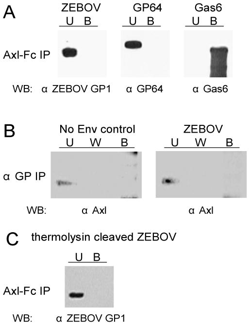
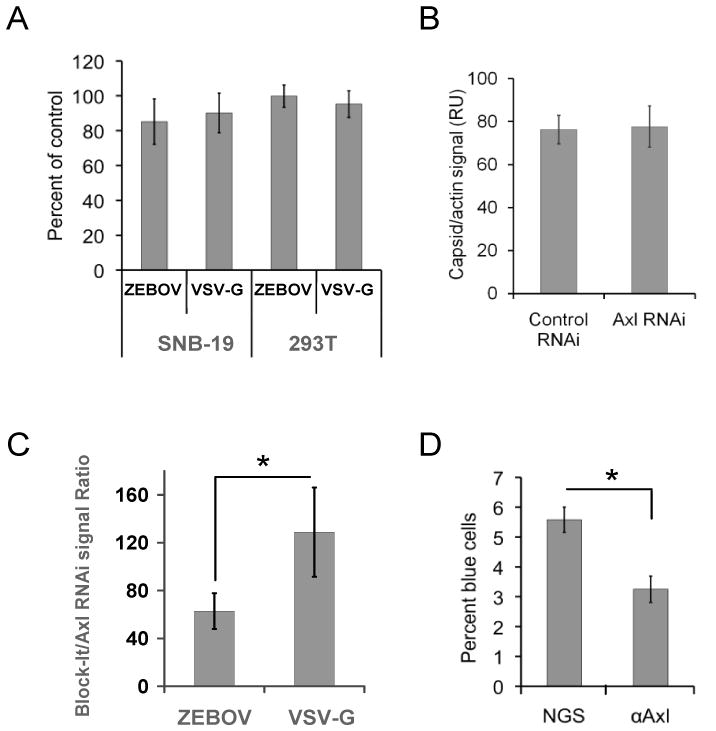
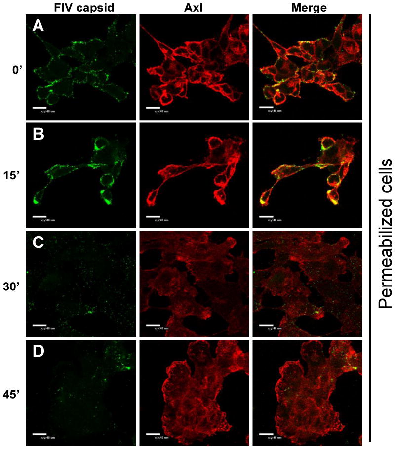
In a bioinformatics-based screen for cellular genes that enhance Zaire ebolavirus (ZEBOV) transduction, AXL mRNA expression strongly correlated with ZEBOV infection. A series of cell lines and primary cells were identified that require Axl for optimal ZEBOV entry. Using one of these cell lines, we identified ZEBOV entry events that are Axl-dependent. Interactions between ZEBOV-GP and the Axl ectodomain were not detected in immunoprecipitations and reduction of surface-expressed Axl by RNAi did not alter ZEBOV-GP binding, providing evidence that Axl does not serve as a receptor for the virus. However, RNAi knock down of Axl reduced ZEBOV pseudovirion internalization and α-Axl antisera inhibited pseudovirion fusion with cellular membranes. Consistent with the importance of Axl for ZEBOV transduction, Axl transiently co-localized on the surface of cells with ZEBOV virus particles and was internalized during virion transduction. In total, these findings indicate that endosomal uptake of filoviruses is facilitated by Axl.


Figures

Fig. 1
Fig. 1. Correlation of ZEBOV-GP pseudotyped VSV transduction with AXL expression in the NCI-60 panel of human tumor cells
A) Relative expression of AXL mRNA expression in the 52 cell lines analyzed in the screen. B) ZEBOV-GP ΔO VSV transduction of the 52 lines. C) VSV-G VSV transduction of the same lines. AXL expression and ZEBOV-GP ΔO-dependent transduction, but not VSV-G-dependent transduction were positively correlated in our transduction screen with a Pearson correlation coefficient (PCC) of 0.517 (p<0.0001). Data shown are the mean of three independent sets of transductions. Gene array data from the individual arrays are available at http://dtp.nci.nih.gov/mtargets/download.html. The data set compared in this study was GC11900. Leuk = leukemia lines; NSCLC = non-small cell lung carcinoma; CNS = central nervous system; P = prostate.
Fig. 2
Fig. 2. Cell surface Axl expression
A) Axl surface expression on two NCI-60 cell lines that were poorly transduced with ZEBOV-GP ΔO VSV and displayed low levels of AXL expression in NCI-60 gene array studies. B) Axl surface expression on two NCI-60 cell lines that were highly transduced with ZEBOV-GP ΔO VSV and displayed robust levels of AXL mRNA expression on the gene arrays. C) Axl expression on the surface of two primary human cell populations, human foreskin fibroblasts (Hff) and umbilical cord endothelial cells (HuVEC). Cells stained with normal goat sera are shown in grey histograms, whereas cells stained with goat anti-human Axl antisera are shown in black histograms.
Fig. 3
Fig. 3. The impact of Axl on ZEBOV transduction is cell dependent
A) Ability of Axl antisera to block ZEBOV-GP- and VSV G-dependent transduction. Cells were incubated with 8 μg/mL of goat anti-Axl sera or normal goat serum (GS) at 4ºC for 30 minutes. VSV-G or ZEBOV-GP ΔO pseudotyped VSV (MOI =0.5) was added to cells and shifted to 37ºC. Twenty hours following transduction, the cells were analyzed for EGFP expression using flow cytometry. Relative transduction values in the presence of antisera are shown as the percent of transduction in the absence of antisera (control values) for each cell population. Shown are the mean and standard error of the mean of three independent experiments. *, p <0.05; **, p < 0.001. B) Ability of Axl antisera to reduce infection of recombinant ZEBOV-GP ΔO VSV. SNB-19 cells were pre-incubated with either 2 μg/mL of anti-Axl antisera or normal goat sera (GS) for 15 minutes prior to addition of virus (MOI= 0.1). Cells were assessed for EGFP expression on days noted. Shown are the mean and standard error of the mean of one experiment that is representative of three independent studies that were performed. C) Ability of Axl expression to increase ZEBOV-GP ΔO pseudotyped VSV entry in the poorly permissive NCI-H522 cell line. NCI-H522 cells were transfected with empty plasmid or plasmid expressing Axl and transduced with VSV pseudotypes (MOI 0.005) 48 h following transfection. Cells were analyzed for EGFP expression 24 h following transduction by flow cytometry. A ratio of transduction relative to transduction in the presence of empty plasmid are shown. Shown are the mean and standard error of the mean of three independent experiments. **, p < 0.001
Fig. 4
Fig. 4. Thermolysin-treated ZEBOV-GP pseudovirions remain sensitive to AXL RNAi treatment
A and B) Knockdown of Axl expression in SNB-19 cells by validated AXL siRNA (Invitrogen) at 48 hours following transfection as assessed by immunoblotting of cell lysates for Axl (A) or cell surface expression of Axl (B). Shown in the black histogram are cells treated with AXL RNAi; the grey histogram represents cells treated with an irrelevant RNAi. C) Thermolysin treated full length ZEBOV-GP FIV pseudovirions remain sensitive to AXL RNAi treatment. Virions were treated with thermolysin or mock treated and added to SNB-19 cells that were treated with either AXL RNAi or an irrelevant RNAi. Cells were evaluated at 48 hours following transduction for β-gal expression. The findings are shown as the ratio of the number of transduced cells found in each treatment divided by control values. The numbers in parenthesis represent the effect of AXL RNAi treatment on transduction relative to the respective control. **, p <0.001.
Fig. 5
Fig. 5. Direct interactions between ZEBOV-GP and Axl cannot be detected
A) Axl-Fc (0.5 μg) was bound to Protein A sepharose beads and incubated with ZEBOV-GP ΔO or baculovirus GP64 pseudotyped FIV particles. Pull down of Gas6 (0.5 μg) served as a positive control in this study. U, unbound; B, Axl-Fc bound. B) ZEBOV pseudotyped particles were bound to protein G beads with anti-ZEBOV-GP1 antisera. SNB-19 cell lysates were passed over the beads and the unbound (U), wash (W), and bound (B) fractions were examined for Axl by immunoblot. C) Axl-Fc does not interact with thermolysin-cleaved ZEBOV FIV particles.
Fig. 6
Fig. 6. Axl is required for post-binding events
A) Soluble Axl-Fc does not interfere with ZEBOV-GP ΔO pseudotyped VSV transduction. ZEBOV and VSV-G virions were pre-incubated with Axl-Fc (50 μg/mL) and transduced on to Axl dependent SNB-19 cells or the Axl-independent 293T cells. Transduction was evaluated by EGFP expression in the transduced populations at 24 hours. Results are shown as the number of cells transduced in the presence of Axl-Fc divided by the number of transduced cells in the absence of treatment. B) RNAi knock down of Axl had no effect on ZEBOV-GP ΔO pseudovirion binding. Forty-eight hours following transfection of AXL RNAi or an irrelevant RNAi into SNB-19 cells, equivalent quantities of ZEBOV-GP ΔO FIV were incubated with cells for one hour at 4°C. Unbound virus was removed and cells were lysed. Lysates were immunoblotted for FIV capsid and quantitated as described in the Materials and Methods. Shown is average pixel values of FIV capsid on the immunoblot divided by the average pixel values for cellular β-actin from 10 independent experiments. C) ZEBOV-GP ΔO FIV internalization, but not VSV-G FIV is decreased in AXL siRNA-treated cells. SNB-19 cells were transfected with AXL siRNA or an irrelevant control siRNA. At 48 hours, ZEBOV-GP ΔO FIV pseudovirions were bound to the cells for 1 hour at 4°C. Unbound virus was removed and cells were shifted to 37°C for 2 hour. Cells were lysed and immunoblotted for FIV capsid and cellular actin. The capsid signal was normalized for actin levels and data are reported as the ratio of the FIV signal in the Axl knock down cells divided by the FIV signal in the cells transfected with an irrelevant RNA. D) Ability of Axl antisera to block ZEBOV VLP fusion events. ZEBOV-GP ΔO-VLPs containing Src-β-lactamase were transduced into SNB-19 cells in the presence or absence of 1:20 dilution of polyclonal antisera against the ectodomain of Axl or normal goat sera (NGS). Entry of β-lactamase into the cytoplasm of cells was evaluated by flow cytometry following incubation of the cells with the fluorescent β-lactamase substrate CCF2/AM for 2 hours. *p < 0.05.
Fig. 7
Fig. 7. Axl transiently associated with ZEBOV-GP ΔO pseudovirions on the cell surface and is internalized as virions enter cells
Confocal microscopy of Axl and FIV capsid during ZEBOV-GP ΔO FIV transduction. A) Localization of Axl and FIV capsid expression in SNB-19 cells incubated with ZEBOV-GP ΔO FIV (MOI=250) for 1 hour at 4°C. B–D) Localization of Axl and FIV capsid in SNB-19 cells following incubation of ZEBOV-GP ΔO FIV (MOI=250) for 1 hour at 4°C, removal of unbound virions and incubation of cells with pre-warmed media at 37°C for 15 minutes (B), 30 minutes (C) or 45 minutes (D). All coverslips were fixed with 2% paraformaldehyde, permeabilized with 0.2% Triton X-100 and immunostained with goat anti-Axl and mouse anti-FIV capsid. Findings shown in panels are representative experiments performed three independent times.
Fig. 8
Fig. 8. Both Axl and ZEBOV-GP ΔO pseudovirions are internalized from the cell surface during infection
Confocal microscopy of Axl and FIV capsid during ZEBOV-GP ΔO FIV transduction in non-permeabilized cells. A) Localization of Axl and FIV capsid expression in SNB-19 cells incubated with ZEBOV-GP ΔO FIV (MOI=250) for 1 hour at 4°C. B–D) Localization of Axl and FIV capsid in SNB-19 cells following incubation of ZEBOV-GP ΔO FIV (MOI=250) for 1 hour at 4°C, removal of unbound virions and incubation of cells with pre-warmed media at 37°C for 15 minutes (B), 30 minutes (C) or 45 minutes (D). All coverslips were fixed with 2% paraformaldehyde and immunostained with goat anti-Axl and mouse anti-FIV capsid. Findings shown in panels are representative experiments performed three independent times.

References

    1. Allavena P, Chieppa M, Monti P, Piemonti L. From pattern recognition receptor to regulator of homeostasis: the double-faced macrophage mannose receptor. Crit Rev Immunol. 2004;24:179–192. - PubMed
    1. Alvarez CP, Lasala F, Carrillo J, Muniz O, Corbi AL, Delgado R. C-type lectins DC-SIGN and L-SIGN mediate cellular entry by Ebola virus in cis and in trans. J Virol. 2002;76:6841–6844. - PMC - PubMed
    1. Baribaud F, Doms RW, Pohlmann S. The role of DC-SIGN and DC-SIGNR in HIV and Ebola virus infection: can potential therapeutics block virus transmission and dissemination? Expert Opin Ther Targets. 2002;6:423–431. - PubMed
    1. Bhattacharyya S, Warfield KL, Ruthel G, Bavari S, Aman MJ, Hope TJ. Ebola virus uses clathrin-mediated endocytosis as an entry pathway. Virology. 2010;401:18–28. - PMC - PubMed
    1. Braunger J, Schleithoff L, Schulz AS, Kessler H, Lammers R, Ullrich A, Bartram CR, Janssen JW. Intracellular signaling of the Ufo/Axl receptor tyrosine kinase is mediated mainly by a multi-substrate docking-site. Oncogene. 1997;14:2619–2631. - PubMed

Publication types

MeSH terms