Review
. 2011 Jul;50(1):1-8.
doi: 10.1016/j.ceca.2011.02.007. Epub 2011 Apr 29.

Analysis of CaM-kinase signaling in cells

Affiliations
Review

Analysis of CaM-kinase signaling in cells

Gary A Wayman et al. Cell Calcium. 2011 Jul.

Abstract

A change in intracellular free calcium is a common signaling mechanism that modulates a wide array of physiological processes in most cells. Responses to increased intracellular Ca(2+) are often mediated by the ubiquitous protein calmodulin (CaM) that upon binding Ca(2+) can interact with and alter the functionality of numerous proteins including a family of protein kinases referred to as CaM-kinases (CaMKs). Of particular interest are multifunctional CaMKs, such as CaMKI, CaMKII, CaMKIV and CaMKK, that can phosphorylate multiple downstream targets. This review will outline several protocols we have used to identify which members and/or isoforms of this CaMK family mediate specific cellular responses with a focus on studies in neurons. Many previous studies have relied on a single approach such as pharmacological inhibitors or transfected dominant-negative kinase constructs. Since each of these protocols has its limitations, that will be discussed, we emphasize the necessity to use multiple, independent approaches in mapping out cellular signaling pathways.


Figures

Figure 1
Figure 1
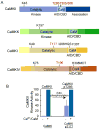
Subunit structure and regulation of CaMKs. A. Schematic diagrams of CaMKs with key residues involved in their regulation by phosphorylation (red font) or that are mutated in making dominant-negative or constitutively-active constructs (black font). See text for details. The ∇ in CaMKK at D434 indicates the site of truncation for the constitutively-active construct. AID, autoinhibitory domain; CBD, calmodulin-binding domain. B-D. Regulation of CaMKs by phosphorylation. Autophosphorylation of Thr286 in CaMKII (B) generates Ca2+/CaM-independent activity (30-70% of total) whereas phosphorylation of the activation loop sites in CaMKI (C) or CaMKIV (D) by CaMKK primarily increases total activity (i.e., with Ca2+/CaM) although CaMKIV exhibits some (~ 10-20%) independent activity.
Figure 2
Figure 2
Immunocytochemical analysis of CaMKII activation in hippocampal neurons is blocked by the CaMKII inhibitor CaMKIIN. Low-density hippocampal cultures were transfected on day 5 with plasmid encoding EGFP-CaMKIIN. The neurons were cultured an additional 9 d and then stained with antibody specific for phospho-Thr286 in CaMKII (pCaMKII), either with (right panels) or without (left panels) prior stimulation with 90 mM KCl (5 min). After KCl stimulation (right panel), nontransfected neurons (arrowheads) show enhanced staining for activated CaMKII (autophosphorylated at Thr286) throughout their cell bodies and processes (bottom panel). EGFP-CaMKIIN-expressing neurons (arrows) show no increase in anti-phospho-CaMKII staining over levels seen in unstimulated cells, demonstrating that overexpression of CaMKIIN inhibited activation of endogenous CaMKII. Scale bar; B, 50 μm. Reproduced from Wayman et al. 2004 by permission from the Journal of Neuroscience.
Figure 3
Figure 3
Dominant-negative CaMKIV distributes throughout the cytoplasm of transfected neurons. A. Low-density hippocampal cultures were transfected on day 3 with plasmid encoding EGFP-dnCaMKIV (upper panels) or a construct modified by addition of a nuclear localization signal, dnCaMKIVnuc (lower panels). Low magnification confocal images of the EGFP signal are shown in the left panels. While the dnCaMKIV lacking a nuclear targeting signal filled the cytoplasm of the cell body (arrow), the whole length of the axon and dendrites, it was largely excluded from the nucleus (right panel). In contrast, EGFP-dnCaMKIVnuc (lower panels) was found exclusively in the nucleoplasm of the transfected neuron (arrow). Scale bars: 50 μm and 5 μm. B. Nuclear restricted dnCaMKIVnuc inhibits NMDA-stimulated CRE-mediated transcription. Hippocamapal neurons (10 DIV) were transfected with a CRE-regulated luciferase reporter, a β-actin promoter-driven β-galactosidase reporter, and a 10-fold excess of empty vector or dnCaMKIVnuc. After 48 hours the neurons were stimulated with 5 or 10 μM NMDA in the presence of 1 μM glycine for 5 hours and assayed for luciferase activity. β-galactosidase assay was conducted using the Galacton substrate (Tropix) according to the manufacturers instructions. The data (±SEM) are expressed as the ratio of luciferase to galactosidase (n = 4).
Figure 4
Figure 4
pCAGGS efficiently expresses dendritic morphology markers in hippocampal neurons. DIV7 hippocampal neurons were transfected with pCAGGS-mRFP-β-Actin and pCAGGSEGFP- Map2B. On DIV12 the neurons were fixed and imaged by confocal microscopy. EGPFMap2B highlights the dendritic arbor whereas mRFP-β-Actin highlights the dendritic spines. Neither of these markers showed detectable expression in glia.

References

    1. Berridge MJ, Lipp P, Bootman MD. Signal transduction. The calcium entry pas de deux. Science. 2000;287:1604–5. - PubMed
    1. Higley MJ, Sabatini BL. Calcium signaling in dendrites and spines: practical and functional considerations. Neuron. 2008;59:902–13. - PubMed
    1. Gaertner TR, Kolodziej SJ, Wang D, Kobayashi R, Koomen JM, Stoops JK, Waxham MN. Comparative analyses of the three-dimensional structures and enzymatic properties of alpha, beta, gamma and delta isoforms of Ca2+-calmodulin-dependent protein kinase II. J Biol Chem. 2004;279:12484–94. - PubMed
    1. Rosenberg OS, Deindl S, Comolli LR, Hoelz A, Downing KH, Nairn AC, Kuriyan J. Oligomerization states of the association domain and the holoenyzme of Ca2+/CaM kinase II. FEBS J. 2006;273:682–94. - PubMed
    1. Haribabu B, Hook SS, Selbert MA, Goldstein EG, Tomhave ED, Edelman AM, Snyderman R, Means AR. Human calcium-calmodulin dependent protein kinase I: cDNA cloning, domain structure and activation by phosphorylation at threonine-177 by calcium-calmodulin dependent protein kinase I kinase. EMBO J. 1995;14:3679–86. - PMC - PubMed

Publication types

MeSH terms

Substances