

Review
doi: 10.1016/j.ceca.2011.03.011.
Epub 2011 May 6.
The endo-lysosomal system as an NAADP-sensitive acidic Ca(2+) store: role for the two-pore channels
Affiliations
- PMID: 21529939
- PMCID: PMC3160778
- DOI: 10.1016/j.ceca.2011.03.011
Item in Clipboard
Review
The endo-lysosomal system as an NAADP-sensitive acidic Ca(2+) store: role for the two-pore channels
Cell Calcium.
2011 Aug.
Abstract
Accumulating evidence suggests that the endo-lysosomal system provides a substantial store of Ca(2+) that is tapped by the Ca(2+)-mobilizing messenger, NAADP. In this article, we review evidence that NAADP-mediated Ca(2+) release from this acidic Ca(2+) store proceeds through activation of the newly described two-pore channels (TPCs). We discuss recent advances in defining the sub-cellular targeting, topology and biophysics of TPCs. We also discuss physiological roles and the evolution of this ubiquitous ion channel family.
2011 Elsevier Ltd. All rights reserved.
Figures
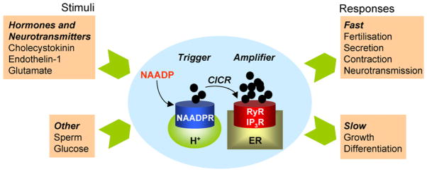
NAADP-mediated Ca2+ signalling. Schematic depicting selected NAADP-linked stimuli (left) and NAADP-regulated Ca2+-dependent cellular responses (right). The intervening diagram summarizes the “trigger” hypothesis for NAADP action, whereby NAADP produced in response to cellular stimulation activates NAADP-sensitive Ca2+ channels located on acidic Ca2+ stores. The resulting local Ca2+ signal is then amplified by neighbouring IP3 and ryanodine receptors on the ER by Ca2+-induced Ca2+ release (CICR) to mediate a larger global Ca2+ signal.
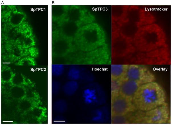
Sub-cellular distribution of sea urchin TPCs. Confocal images of Stronglylocentrotus purpuratus embryos (19 h post fertilisation) that had been injected with mRNA encoding for GFP-tagged SpTPC1 (A, top), SpTPC2 (A, bottom) and SpTPC3 (B). In B, the embryos were counterstained with lysotracker red (red) and Hoechst (blue) to label acidic organelles and nuclei, respectively. An overlay of the images is shown in the bottom right panel. All scale bars = 5 μm.
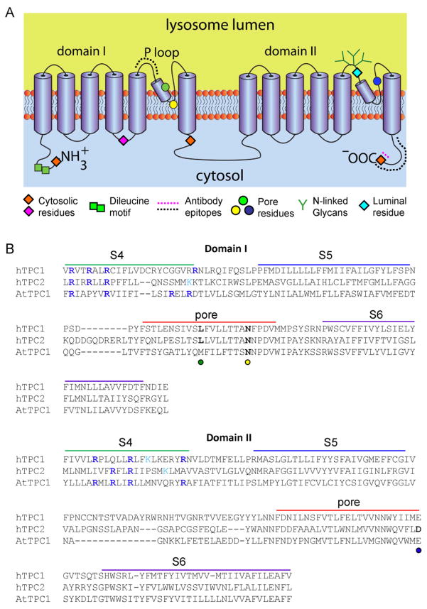
Structural and biophysical properties of TPCs. (A) Proposed topology of TPCs. TPCs are predicted to contain two repeated domains each comprising 6 trans-membrane regions and a putative pore-loop between the fifth and sixth transmembrane regions. Both termini and the loop connecting the domains are predicted to be cytosolic. Positions at which fluorophores were introduced and their accessibility to trypsin assessed in fluorescence protease protection assays [58] are shown by diamonds. The results confirmed the positions of the indicated regions to either the cytosol (orange for both HsTPC1 and HsTPC2; pink for HsTPC1) or lumen (cyan; HsTPC1). An N-terminal dileucine endo-lysosomal targeting motif is shown by green squares. The dotted lines represent the epitopes in TPC1 (pink) and TPC2 (black) to which antibodies used for topology mapping were raised [58]. Pore residues are highlighted by circles (see B for further detail). Green branches indicate N-linked glycosylation sites within the pore loop of domain II [47,58]. (B) Multiple sequence alignment (using ClustalW2) of putative pore regions of human TPC1 (HsTPC1, NP 001137291.1) and TPC2 (HsTPC2, NP 620714.2), and of Arabidopsis TPC1 (AtTPC1, NP 567258.1) from domains I and II. Possible positions of the S4–S6 membrane-spanning regions are from [38]. For voltage-gated cation channels, positively charged residues (usually arginine) aligned on one side of the S4 helix provide the voltage-sensor. Residues that might provide such an arrangement in TPC channels are highlighted in blue (arginine) and cyan (lysine) in each of the S4 regions. There are fewer such residues in HsTPC2 than in HsTPC1 and AtTPC1. Mutation of a conserved Leu (L273 for HsTPC1 and L265 for HsTPC2, green circle) [41,43] or Asn (N257 for mouse TPC2, equivalent to N273 of HsTPC2, yellow circle) [69] within the putative P-loop of domain I affect either Ca2+ release, conductance or ion selectivity, consistent with their proposed positions within the pore. A conserved acidic residue (D660 for HsTPC2 and E643 for mTPC2, blue circle) within the proposed P loop of domain II also appears to be critical for channel function [69], suggesting that the second putative P loop also contributes to formation of the pore.
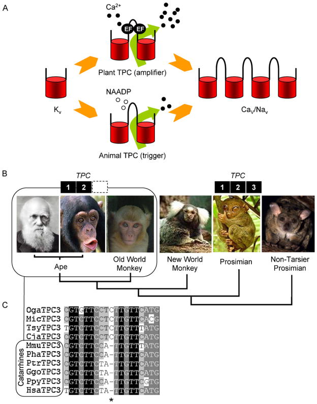
Evolution of TPCs. (A) Schematic outlining a possible trajectory for the evolution of voltage-gated ion channels and TPCs. Voltage-sensitive K+ channels (Kv; one-repeat) may have duplicated to form TPCs (two-repeat) and the TPCs duplicated to form voltage-sensitive Ca2+ (Cav) and Na+ (Nav) channels (four-repeat). Plant TPCs by virtue of their EF hands may serve to amplify Ca2+ signals (middle top) whereas animal TPCs (which lack EF hands) may have evolved to trigger Ca2+ signals in response to NAADP (middle bottom). (B) TPC gene complement of selected primates (from left to right; Homo sapiens, Pan troglodytes, Maccaca mulatta, Callithrix jacchus, Tarsius syrichta, Otolemur garnettii) highlighting lack of a functional TPC3 gene in Catarrhines (box). (C) Multiple sequence alignment of primate genomic sequences. A conserved cytosine residue within the TPC3 gene (*) is deleted in Cattarhines [98]. Abbreviations used are: Oga, Otolemur garnettii; Mic, Microcebus murinus; Tsy, Tarsius syrichta; Cja, Callithrix jacchus; Mmu, Maccaca mulatta; Pha, Papio hamadryas; Ptr, Pan troglodytes; Ggo, Gorilla gorilla; Ppy, Pongo pygmaeus; and Hsa, Homo sapiens.
References
-
- Berridge MJ, Lipp P, Bootman MD. The versatility and universality of calcium signalling. Nat Rev Mol Cell Biol. 2000;1:11–21. - PubMed
-
- Rizzuto R, Pozzan T. When calcium goes wrong: genetic alterations of a ubiquitous signaling route. Nat Genet. 2003;34:135–141. - PubMed
-
- Clapham DE. Calcium signaling. Cell. 2007;131:1047–1058. - PubMed
-
- Patel S, Joseph SK, Thomas AP. Molecular properties of inositol 1,4,5-trisphosphate receptors. Cell Calcium. 1999;25:247–264. - PubMed
-
- Taylor CW, Genazzani AA, Morris SA. Expression of inositol trisphosphate receptors. Cell Calcium. 1999;26:237–251. - PubMed
Publication types
MeSH terms
Substances
Grants and funding
LinkOut - more resources
Full Text Sources
Miscellaneous
