Review
. 2011 Jun;140(7):1895-908.
doi: 10.1053/j.gastro.2011.04.038. Epub 2011 Apr 23.

Functions of autophagy in hepatic and pancreatic physiology and disease

Affiliations
Review

Functions of autophagy in hepatic and pancreatic physiology and disease

Mark J Czaja. Gastroenterology. 2011 Jun.

Abstract

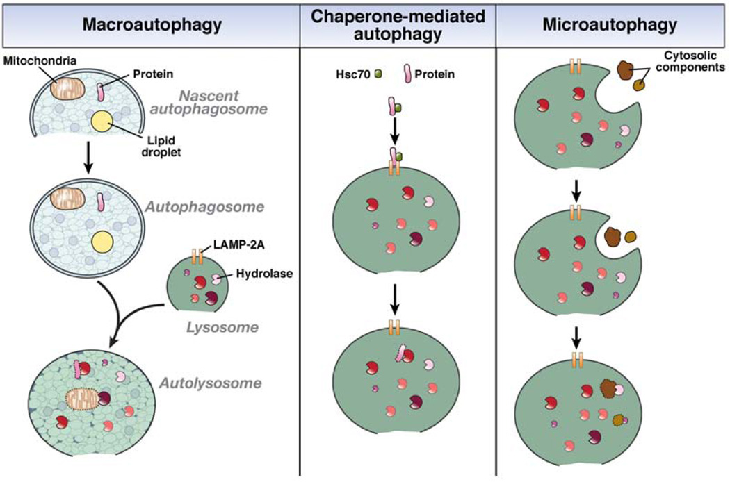
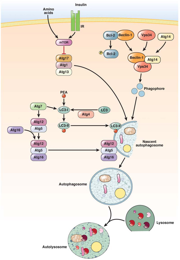
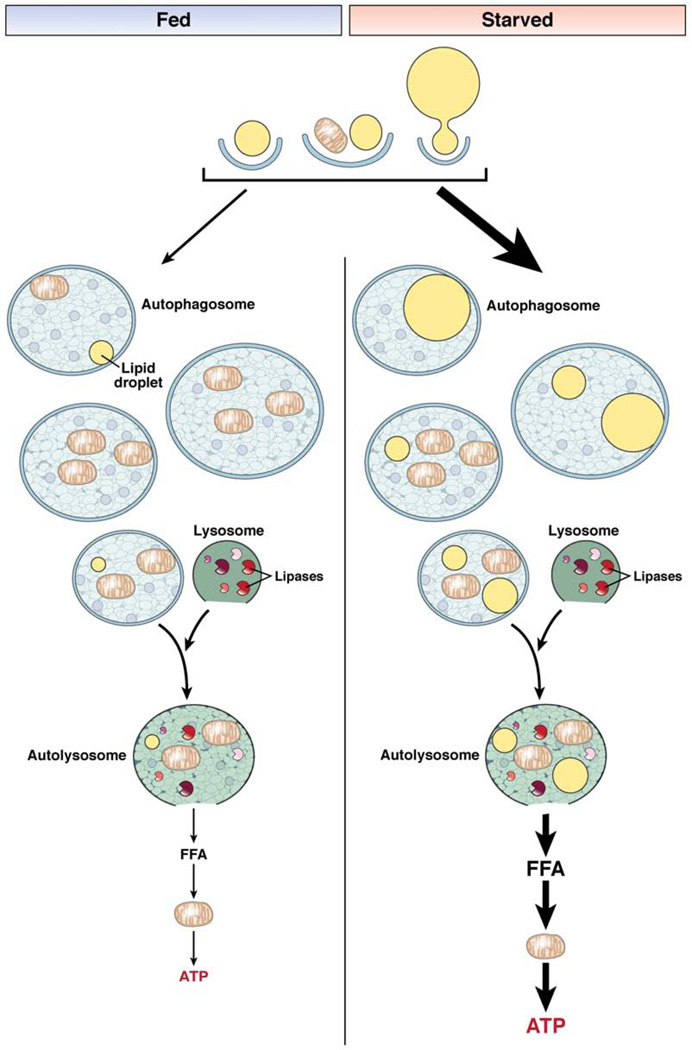
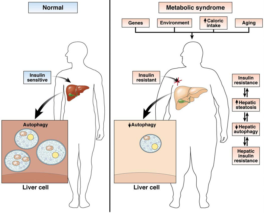
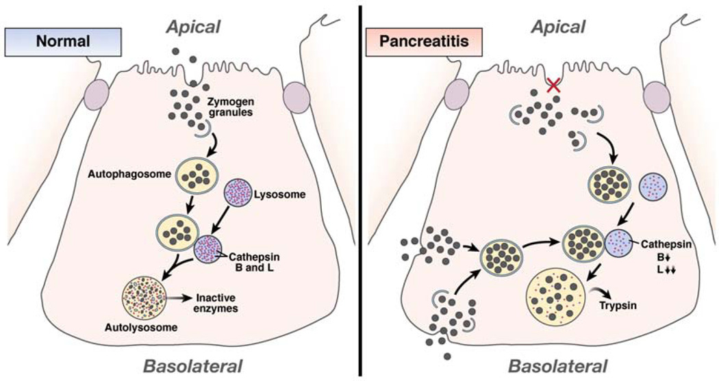
Autophagy is a lysosomal pathway that degrades and recycles intracellular organelles and proteins to maintain energy homeostasis during times of nutrient deprivation and to remove damaged cell components. Recent studies have identified new functions for autophagy under basal and stressed conditions. In the liver and pancreas, autophagy performs the standard functions of degrading mitochondria and aggregated proteins and regulating cell death. In addition, autophagy functions in these organs to regulate lipid accumulation in hepatic steatosis, trypsinogen activation in pancreatitis, and hepatitis virus replication. This review discusses the effects of autophagy on hepatic and pancreatic physiology and the contribution of this degradative process to diseases of these organs. The discovery of novel functions for this lysosomal pathway has increased our understanding of the pathophysiology of diseases in the liver and pancreas and suggested new possibilities for their treatment.


Conflict of interest statement

Conflicts of interest

The author discloses no conflicts.

Figures

Figure 1
Figure 1
The 3 pathways of autophagy .In macroautophagy, doubie membrane of unclear origin forms around cytosolic components such as mitochondria, lipid droplets, and proteins. The membrane elongates to completely enclose the cellular elements within an autophagosome that translocates to a lysosome containing degradative hydrolases. The 2 structures fuse into an autolysosome, in which the cellular components are degraded by the lysosomal hydrolases. In CMA, cytosolic proteins with a specific pentapeptide motif are recognized by the chaperone Hsc70. This complex binds to the lysosomal LAMP-2A receptor for protein internalization and proteolytic degradation. Microautophagy involves the uptake of cellular components, both organelles and proteins, within an invagination of the lysosomal membrane for enzymatic degradation in the lysosome.
Figure 2
Figure 2
Pathways that control levels of macroautophagy. Macroautophagy is regulated by 3 major pathways. The first is an inhibitory pathway in which nutrient or insulin stimulation of the mTOR signaling pathway blocks autophagosome formation (red line). Two other pathways are stimulatory. In one, phosphorylation of Bcl-2 dissociates it from beclin-1, which allows beclin-1 to form a complex with the PI3K Vps34 that produces phosphatidylinositol 3-phosphate, which is required for induction of autophagy. The other pathway involves a series of conjugation steps that generate LC3-II and the Atg5–Atg12–Atg16 protein complex, which are both necessary for autophagosome formation. IR, insulin receptor; PEA, phosphatidylethanolamine.
Figure 3
Figure 3
Autophagic degradation of lipids. In lipophagy, lipid droplets are sequestered in autophagosomes for delivery to lysosomes for breakdown. Small lipid droplets can be taken up whole by an autophagosome, or alternatively portions of large lipid droplets can be degraded. The autophagosome can contain lipid, as the only substrate, or together with other cellular constituents such as mitochondria. The autophagosome eventually fuses with a hydrolase-containing lysosome whose lipases degrade the lipid. This process results in the release of free fatty acids (FFA) that can be oxidized in mitochondria to supply the cell with ATP. In the fed state, lipid breakdown by autophagy is not needed for nutrient supply and autophagy is suppressed. As a result, movement of lipids into the autophagic pathway is minimal. Most of the autophagosomes contain cargo other than lipid or occasionally lipid mixed with other cellular components. The amount of ATP generated by this process in the fed state is minor. In the starved state, autophagy is induced and selectively targets lipid droplets to utilize lipid stores to supply the cell with energy in the absence of other nutrients. As a result, more autophagosomes contain lipid, either alone or in combination with other cargo, enabling the cell to generate ATP from FFA breakdown to maintain energy homeostasis.
Figure 4
Figure 4
Relationship of altered hepatic autophagy to other manifestations of the metabolic syndrome. Numerous factors can contribute to a decrease in hepatic autophagic function, including genetic and environmental factors and the effects of aging. In individuals with the metabolic syndrome, obesity and resultant peripheral insulin resistance can decrease levels of hepatic autophagy. Obesity, insulin resistance, and defective hepatic autophagy can all act to promote the development of hepatic steatosis and hepatic insulin resistance. Increased lipid content in the liver can then further impair hepatic autophagy, leading to increased insulin resistance and its manifestations in other organs.
Figure 5
Figure 5
Autophagy mediates trypsin activation in pancreatitis. In a normal acinar cell, some trypsinogen-containing zymogen granules that are not secreted from the apical membrane can be taken up by autophagosomes for removal. These autophagosomes fuse with lysosomes to form autolysosomes in which the contents of the granules, including pancreatic enzymes, are degraded by lysosomal enzymes, preventing their release. In pancreatitis, a block in enzyme secretion leads to an accumulation of intracellular zymogen granules, which increases uptake of these structures by autophagosomes. Alternatively or additively, granules improperly released from the basolateral surface of the acinar cell are taken back up by the cell and sequestered by autophagosomes. Following autophagosome-lysosome fusion, content degradation fails to occur because of decreased levels of cathepsin B and L. The more pronounced defect in cathepsin L, which inactivates trypsin, than in the activating enzyme cathepsin B leads to trypsin generation within the autolysosome. Active enzymes are released from these enlarged autolysosomes into the cytoplasm to initiate pancreatic cellular injury.

References

    1. Finn PF, Dice JF. Proteolytic and lipolytic responses to starvation. Nutrition. 2006;22:830–844. - PubMed
    1. Iwata J, Ezaki J, Komatsu M, et al. Excess peroxisomes are degraded by autophagic machinery in mammals. J Biol Chem. 2006;281:4035–4041. - PubMed
    1. Kim I, Rodriguez-Enriquez S, Lemasters JJ. Selective degradation of mitochondria by mitophagy. Arch Biochem Biophys. 2007;462:245–253. - PMC - PubMed
    1. Cuervo AM, Knecht E, Terlecky SR, et al. Activation of a selective pathway of lysosomal proteolysis in rat liver by prolonged starvation. Am J Physiol. 1995;269:C1200–C1208. - PubMed
    1. Komatsu M, Waguri S, Ueno T, et al. Impairment of starvation-induced and constitutive autophagy in Atg7-deficient mice. J Cell Biol. 2005;169:425–434. - PMC - PubMed