Review
. 2011 Jul;94(2):115-32.
doi: 10.1016/j.pneurobio.2011.04.009. Epub 2011 Apr 22.

Function and mechanism of axonal targeting of voltage-sensitive potassium channels

Affiliations
Review

Function and mechanism of axonal targeting of voltage-sensitive potassium channels

Chen Gu et al. Prog Neurobiol. 2011 Jul.

Abstract

Precise localization of various ion channels into proper subcellular compartments is crucial for neuronal excitability and synaptic transmission. Axonal K(+) channels that are activated by depolarization of the membrane potential participate in the repolarizing phase of the action potential, and hence regulate action potential firing patterns, which encode output signals. Moreover, some of these channels can directly control neurotransmitter release at axonal terminals by constraining local membrane excitability and limiting Ca(2+) influx. K(+) channels differ not only in biophysical and pharmacological properties, but in expression and subcellular distribution as well. Importantly, proper targeting of channel proteins is a prerequisite for electrical and chemical functions of axons. In this review, we first highlight recent studies that demonstrate different roles of axonal K(+) channels in the local regulation of axonal excitability. Next, we focus on research progress in identifying axonal targeting motifs and machinery of several different types of K(+) channels present in axons. Regulation of K(+) channel targeting and activity may underlie a novel form of neuronal plasticity. This research field can contribute to generating novel therapeutic strategies through manipulating neuronal excitability in treating neurological diseases, such as multiple sclerosis, neuropathic pain, and Alzheimer's disease.


Figures

Fig. 1
Fig. 1. Structural diagram of potassium channel α subunits
(A) Structure diagram of the α subunits of three major groups in the potassium channel superfamily. The α subunits in the group I contain six to seven transmembrane (TM) segments and one pore-forming loop. The α subunits in the group II contain two TM segments and one pore-forming loop. The α subunits in the group III contain four TM segments and two pore-forming loops. “N” and “C” indicate the amino and carboxyl termini of the protein, respectively. (B) Structure diagram of the α subunits of voltage-gated K+ (Kv) channels (Kv1 to Kv4), KCNQ (Kv7) channels, and the BK (Slo1) channel.
Fig. 2
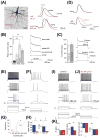
Fig. 2. Regulation of action potential (AP) firing by Kv1 channels localized at axon initial segments
(A) AP is initiated at the AIS of pyramidal neurons. Photograph of a biocytin-filled layer 5 pyramidal neuron indicates the soma, the unmyelinated AIS, a myelinated axonal region, and an axonal bleb site accessible for whole-cell recording (left). Example traces show APs evoked by somatic current injection, and recorded at the AIS 38 μm from the axon hillock (red) and at the soma (black) (top), and recorded at an axon bleb 620 μm from the hillock (bottom). (B) Mean amplitude of slowly inactivating K+ currents evoked by voltage steps from −110 to +45 mV recorded in cell-attached patches from the soma, proximal AIS (5–30 mm from hillock), distal AIS (35–55 mm), and axonal sites (70–400 mm). Asterisks indicate significant increases in K+ peak amplitudes compared to the soma (*** p<0.001 and ** p<0.01). Traces show examples of slow-inactivating K+ current at the indicated locations (averages of four to six patches). (C) Outside-out recordings of K+ currents from AIS or axonal blebs before and after puffs with 500 nM DTX-1. Subtracted traces show the DTX-sensitive current. (D) Impact of bath application of 50–100 nM DTX-1 on somatic (top) and axonal (bottom) APs before (black) and several minutes after DTX-1 (red) ((A)–(D) modified from Kole et al., 2007 with copyright permission from Neuron). (E) Discharge pattern of a fast-spiking (FS) neuron in layer 2/3 barrel cortex. Shown are voltage responses to 600 ms current injections of, from bottom to top, −100, 100, 345, 360, and 440 pA. Gray bars highlight delay to first spike. Inset in the topmost trace shows the first two spikes on an expanded timescale (Scale bar, 10 ms). (F) Firing pattern of an adjacent pyramidal neuron. (G) f–I curves for a subset of representative FS cells (blue) and PCs (red), with the cells in (E) and (F) indicated by dots. (H) Summary data illustrating initial firing frequency and the slope of the f–I relation for FS cells and PCs. (I) Firing pattern of an FS cell in layer 2/3 barrel cortex to current injections. (J) Same cell as in (I), after bath application of 50 nM DTX-1. (K) Summary data. DTX-1 produces no change in Rm or fi but decreased ITH and shifted the voltage threshold for AP generation to hyperpolarized potentials ((E)–(K) modified from Goldberg et al., 2008 with copyright permission from Neuron).
Fig. 3
Fig. 3. Kv1 channels are mainly localized in axon initial segments, juxtaparanodal regions, axonal terminals in nervous systems
(A) Distribution of Nav and Kv1.2 channels in the AIS of pyramidal neurons and their relationship with chandelier cell axonal terminals (modified from Inda et al., 2006 with copyright permission from Proc. Natl. Acad. Sci. U.S.A.) (B) Double labeling by using anti-Kv1.2 (green) and anti-Nav channel (red) antibodies in myelinated rat spinal cord (modified from Rasband and Trimmer, 2001 with copyright permission from J. Comp. Neurol.). Scale bar, 10 μm. (C) Localization of Kv1.1 in mouse cerebellar basket cell terminals. General distribution of Kv1.1 in mouse cerebellum (top) and higher magnification of the boxed region (bottom) (modified from Wang et al., 1994 with copyright permission from the J. Neurosci.) (D) Double labeling reveals segregation of endogenous Kv1.2 and Kv4.2 in mature hippocampal neurons (21 DIV) (modified from Gu et al., 2003 with copyright permission from Science). Immunofluorescence intensity profiles, along a 40-mm line (black, lower left) were measured from the original 16-bit TIFF images with NIH Image J (lower right). AU, arbitrary unit.
Fig. 4
Fig. 4. Mechanisms underlying Kv1 channel targeting in axons
(A) Axonal transport of Kv1 channels. The study using live cell imaging on hippocampal neuron axons expressing YFP-Kv1.2 suggests that Kv1/Kvβ channel complexes reside in small round vesicles moving anterogradely along axons. These vesicles are carried by KIF3A/kinesin II motors, where plus end tracking protein EB1 also plays a critical role. It is also possible that KIF5B might transport Kv1 channels anterogradely under some conditions. Kv1 channel-containing vesicles can also be transported retrogradely, which is potentially mediated by dynein complex. How Kv1 channel complex is linked to dynein remains unknown. (B) Targeting of Kv1 channels into the JXP regions in meylinated axons. TAG-1 expressed on myelinating glial and axonal membranes can form homomultimers in trans. TAG-1 can bind to Caspr2 on axonal membranes in cis. Since both Kv1 channels and Caspr2 have PDZ-binding ligand at their C-termini, PDZ domain-containing proteins, such as PSD-95 and PSD-93, may cluster Kv1 channel complexes and Caspr2 together. Therefore, Kv1, Caspr2 and TAG-1 co-cluster in the juxtaparanodal regions of myelinated axons.
Fig. 5
Fig. 5. Axon-dendrite targeting of Kv3 channels
(A) Differential localization of Kv3.1b and Kv1.2 in axons of central nervous systems. These are images of longitudinal sections of the ventral column (vc) of unfixed lumbar spinal cord from adult mice. In the spinal cord, the narrow band of nodal Kv3.1b staining (top) is flanked by two broader regions of JXP Kv1.2 staining (middle). The merged image is at the bottom. Note the relative lack of labeling for Kv3.1b in the nodes of peripheral nervous systems (double arrowheads) of the ventral rootlet (vr) (modified from Devaux et al., 2003 with copyright permission from J. Neurosci.). (B) Kv3.3 (flexor digitorum brevis)(top) and Kv3.4 (transverses abdominus)(bottom) localize in presynaptic motor nerve terminals revealed by double labeling with the postsynaptic nicotinic acetylcholine receptors by FITC-α bungarotoxin (left). The presynaptic localization of Kv3.3 and Kv3.4 was further confirmed by electron microscopy studies (modified from Brooke et al., 2004 with copyright permission from Eur. J. Neurosci.). (C) Presence of Kv3.3 in soma and dendrites of Purkinje neurons in rat cerebellum (modified from Martina et al., 2003 with copyright permission from J. Neurosci.).
Fig. 6
Fig. 6. Local trafficking and long-distance transport of axonal Kv3.1 channels
A proposed model for the potential interplay of KIF5 and ankyrin G during transportation of Kv3.1 channels through the AIS. (A) A KIF5 motor directly binds to the Kv3.1 T1 tetramer via its C-terminal tail and transports the channel tetramer. Kv3.1 channel may or may not also bind to ankyrin G through its C-terminus assembled together with the T1 tetramer. For clarification, only one C-terminal tail is shown here. (B) Disassembly or conformational change of the T1 tetramer dissociates the channel from KIF5 and allows a stronger binding of ankyrin G to the C-terminus. The process may involve more intermediate states besides the two shown here (modified from Xu et al., 2010 with copyright permission from J. Neurosci.).
Fig. 7
Fig. 7. Axonal localization of KCNQ channels
(A)–(D) Colocalization of KCNQ2 and KCNQ3 in AISs in cortex. These are images of horizontal sections of unfixed mouse brain immunolabeled for KCNQ2, KCNQ3, and ankyrin G; DAPI was used as a nuclear counterstain in (A) and (B). In the CA3 region of the hippocampus, many AISs in the stratum pyramidale (sp), as well as the mossy fibers of the stratum lucidum (sl), are strongly KCNQ2 positive (A). The stratum radiatum (sr) and stratum oriens (so) are indicated. KCNQ3 was found with KCNQ2 in the AISs of some pyramidal cells in CA3 but also in the mossy fibers (B). KCNQ3 colocalized with both ankyrin G and KCNQ2 in the AIS of neurons from the CA1 (C) and temporal neocortex (D). The asterisk in (B) marks a KCNQ3-positive blood vessel. (E) KCNQ2 localized to CNS nodes and initial segments. Anti-KCNQ2 (red), anti-panNav (green, top), and anti-Kv1.2 (green, bottom) antibodies were used to stain rat spinal cord. (F) KCNQ2 is localized to PNS nodes of Ranvier. The staining was performed on unfixed teased fibers from adult rat sciatic nerves (modified from Devaux et al., 2004 with copyright permission from J. Neurosci.).
Fig. 8
Fig. 8. Motifs of KCNQ channels responsible for axonal targeting
A structure diagram of KCNQ subunit shows the important trafficking motifs in the C-terminus. MP domain, membrane proximal domain (amino acids 323–500); A domain, amino acids 501–579; SID, subunit interaction domain (amino acids 580–623); Ankyrin G-binding domain, amino acids 624–844.
Fig. 9
Fig. 9. Axon-dendrite targeting of BK channels
(A) Immunolocalization of BK channels in hippocampus. The monoclonal anti-BK antibody (L6/60) (red) was used to stain the hippocampal sections from wild type mouse (top) and from BK-knockout mouse (bottom). The anti-Kv2.1 staining (green) was used as control. (B)–(D) In rat cerebellar Purkinje cell layer, BK channels are localized in Kv1.2-positive basket cell terminals (B), around the neurofilament-positive AISs (C), and in calbindin-positive Purkinje cell soma and dendrites (D). (E) BK channel staining associates with axons and dendrites of cultured hippocampal neurons (modified from Misonou et al., 2006 with copyright permission from J. Comp. Neurol.).

Similar articles

Cited by

References

    1. Alioua A, Lu R, Kumar Y, Eghbali M, Kundu P, Toro L, Stefani E. Slo1 caveolin-binding motif, a mechanism of caveolin-1-Slo1 interaction regulating Slo1 surface expression. J Biol Chem. 2008;283:4808–4817. - PubMed
    1. Arnold DB, Clapham DE. Molecular determinants for subcellular localization of PSD-95 with an interacting K+ channel. Neuron. 1999;23:149–157. - PubMed
    1. Asbury CL, Fehr AN, Block SM. Kinesin moves by an asymmetric hand-over-hand mechanism. Science. 2003;302:2130–2134. - PMC - PubMed
    1. Atzori M, Lau D, Tansey EP, Chow A, Ozaita A, Rudy B, McBain CJ. H2 histamine receptor-phosphorylation of Kv3.2 modulates interneuron fast spiking. Nat Neurosci. 2000;3:791–798. - PubMed
    1. Barry J, Gu Y, Gu C. Polarized targeting of L1-CAM regulates axonal and dendritic bundling in vitro. Eur J Neurosci. 2010;32:1618–1631. - PMC - PubMed

Publication types

MeSH terms

Substances