Review
. 2011 Jul;94(2):133-48.
doi: 10.1016/j.pneurobio.2011.04.011. Epub 2011 Apr 22.

Control of synapse development and plasticity by Rho GTPase regulatory proteins

Affiliations
Review

Control of synapse development and plasticity by Rho GTPase regulatory proteins

Kimberley F Tolias et al. Prog Neurobiol. 2011 Jul.

Abstract

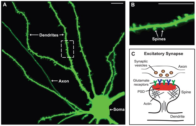
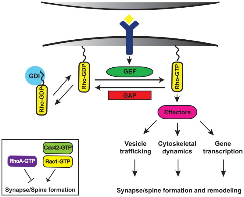
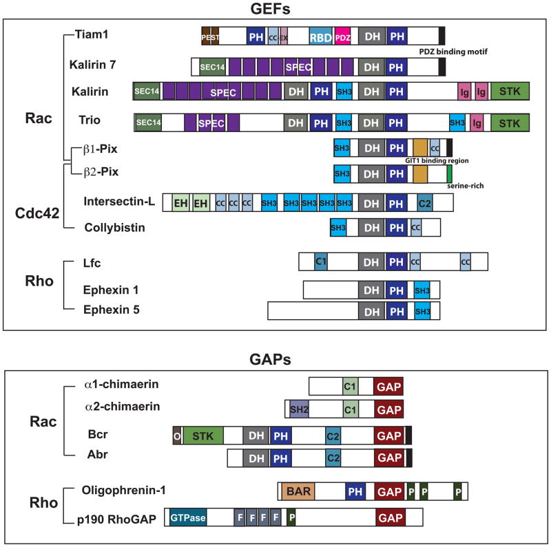
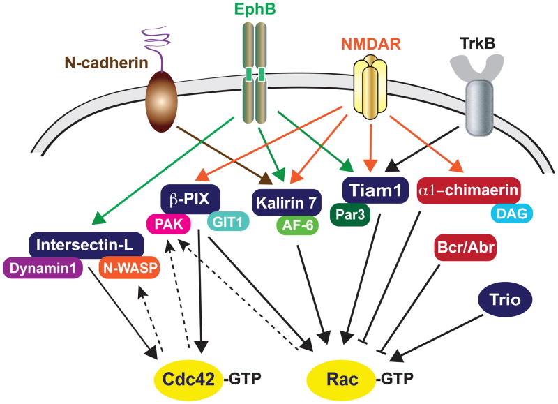
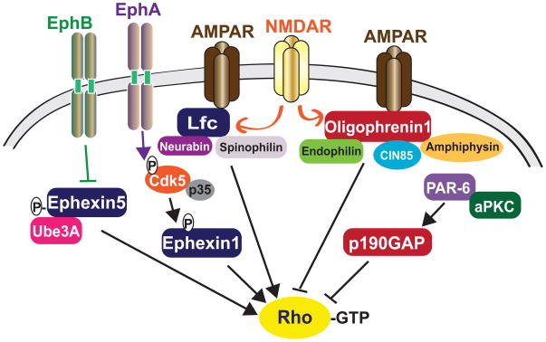
Synapses are specialized cell-cell contacts that mediate communication between neurons. Most excitatory synapses in the brain are housed on dendritic spines, small actin-rich protrusions extending from dendrites. During development and in response to environmental stimuli, spines undergo marked changes in shape and number thought to underlie processes like learning and memory. Improper spine development, in contrast, likely impedes information processing in the brain, since spine abnormalities are associated with numerous brain disorders. Elucidating the mechanisms that regulate the formation and plasticity of spines and their resident synapses is therefore crucial to our understanding of cognition and disease. Rho-family GTPases, key regulators of the actin cytoskeleton, play essential roles in orchestrating the development and remodeling of spines and synapses. Precise spatio-temporal regulation of Rho GTPase activity is critical for their function, since aberrant Rho GTPase signaling can cause spine and synapse defects as well as cognitive impairments. Rho GTPases are activated by guanine nucleotide exchange factors (GEFs) and inhibited by GTPase-activating proteins (GAPs). We propose that Rho-family GEFs and GAPs provide the spatiotemporal regulation and signaling specificity necessary for proper Rho GTPase function based on the following features they possess: (i) existence of multiple GEFs and GAPs per Rho GTPase, (ii) developmentally regulated expression, (iii) discrete localization, (iv) ability to bind to and organize specific signaling networks, and (v) tightly regulated activity, perhaps involving GEF/GAP interactions. Recent studies describe several Rho-family GEFs and GAPs that uniquely contribute to spinogenesis and synaptogenesis. Here, we highlight several of these proteins and discuss how they occupy distinct biochemical niches critical for synaptic development.


Figures

Figure 1
Figure 1. Dendritic spines are the primary sites of excitatory synapses in the brain
A. Shown is an example of a rat hippocampal neuron expressing green fluorescent protein (GFP). Neurons possess a soma, an axon and branched dendrites containing dendritic spines. B. Image shows an enlargement of the dashed box pictured in A, which provides a clearer view of spines. C. Schematic of an excitatory synapse, which forms between a dendritic spine and a presynaptic bouton on an axon. The postsynaptic density (PSD), which contains glutamate receptors, scaffolding proteins and other signaling molecules, is located on the spine head. Spines are also enriched in actin filaments. Scale bar: 10 νm
Figure 2
Figure 2. Model of Rho GTPase signaling at synapses
Rho GTPases function as binary switches by cycling between an active, GTP-bound form and an inactive, GDP-bound form. Rho GTPase activity is tightly regulated in space and time by three different classes of regulatory proteins: guanine nucleotide exchange factors (GEFs), GTP-activating proteins (GAPs), and guanine nucleotide dissociation inhibitors (GDIs). In their active state, Rho GTPases interact with downstream effectors that regulate a variety of cellular processes, which ultimately contribute to spine morphogenesis and excitatory synapse development. The Rho GTPases Rac and Cdc42 promote the formation and growth of synapses and spines, whereas RhoA inhibits synapse development.
Figure 3
Figure 3. Domain structures of synaptic Rho-family GEFs and GAPs
Shown are the domain structures of proteins mentioned in this review. The following abbreviations are used in this figure: PH: pleckstrin homology domain, CC-Ex: coiled coil-extended region, RBD: Ras-binding domain, PDZ: domain in PSD-95, Dlg, and ZO-1/2, DH: Dbl homology domain, SEC14: domain in phosphatidylinositol transfer protein Sec14, SPEC: spectrin-like repeats, SH3: Src homology 3 domain, EH: Eps15 homology domain, C2: protein kinase C conserved region 2, C1: protein kinase C conserved region 1, BAR: Bin/Amphiphysin/Rvs domain, and P: proline-rich regions.
Figure 4
Figure 4. Regulation of Rac/Cdc42 signaling at synapses
The Rac/Cdc42-GEFs Tiam1, Kalirin7, β-PIX, Intersectin-L and Trio promote Rac/Cdc42 activation, whereas the Rac-GAPs α1-chimaerin, Bcr and Abr inhibit Rac activation at synapses. Although these GEFs and GAPs are regulated by common upstream receptors, e.g. NMDA receptors, EphB, TrkB and N-cadherins, the unique role of each regulator likely arises from the networks of interacting proteins that are unique to each GEF or GAP and/or differential upstream regulation of these molecules in a temporal and spatial manner. GEFs are in blue and GAPs are in red, and dashed lines represent positive feedback loops.
Figure 5
Figure 5. Regulation of RhoA signaling at synapses
The RhoA-GEFs Lfc, Ephexin1 and Ephexin5 activate RhoA activity, whereas the RhoA-GAPs Oligophrenin1 and p190 RhoGAP inhibit RhoA activity. These regulatory proteins are controlled by upstream receptors, such as NMDA receptors, AMPA receptors, and Eph receptors. As is the case for Rac1/Cdc42 GEFs and GAPs, RhoA regulatory proteins likely achieve their specific functions through a unique set of interactions with downstream effectors and other proteins. GEFs are in blue and GAPs are in red.

References

    1. Ahmadian G, Ju W, Liu L, Wyszynski M, Lee SH, Dunah AW, Taghibiglou C, Wang Y, Lu J, Wong TP, Sheng M, Wang YT. Tyrosine phosphorylation of GluR2 is required for insulin-stimulated AMPA receptor endocytosis and LTD. Embo J. 2004;23:1040–1050. - PMC - PubMed
    1. Ahmed S, Kozma R, Monfries C, Hall C, Lim HH, Smith P, Lim L. Human brain n-chimaerin cDNA encodes a novel phorbol ester receptor. Biochem J. 1990;272:767–773. - PMC - PubMed
    1. Ahmed S, Lee J, Kozma R, Best A, Monfries C, Lim L. A novel functional target for tumor-promoting phorbol esters and lysophosphatidic acid. The p21rac-GTPase activating protein n-chimaerin. J Biol Chem. 1993;268:10709–10712. - PubMed
    1. Alexander M, Chan KK, Byrne AB, Selman G, Lee T, Ono J, Wong E, Puckrin R, Dixon SJ, Roy PJ. An UNC-40 pathway directs postsynaptic membrane extension in Caenorhabditis elegans. Development. 2009;136:911–922. - PubMed
    1. Allen KM, Gleeson JG, Bagrodia S, Partington MW, MacMillan JC, Cerione RA, Mulley JC, Walsh CA. PAK3 mutation in nonsyndromic X-linked mental retardation. Nat Genet. 1998;20:25–30. - PubMed

Publication types

MeSH terms

Substances