

What is the best way to contour lung tumors on PET scans? Multiobserver validation of a gradient-based method using a NSCLC digital PET phantom
- PMID: 21531085
- PMCID: PMC3877699
- DOI: 10.1016/j.ijrobp.2010.12.055
What is the best way to contour lung tumors on PET scans? Multiobserver validation of a gradient-based method using a NSCLC digital PET phantom
Abstract
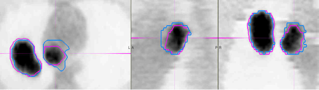
Purpose: To evaluate the accuracy and consistency of a gradient-based positron emission tomography (PET) segmentation method, GRADIENT, compared with manual (MANUAL) and constant threshold (THRESHOLD) methods.
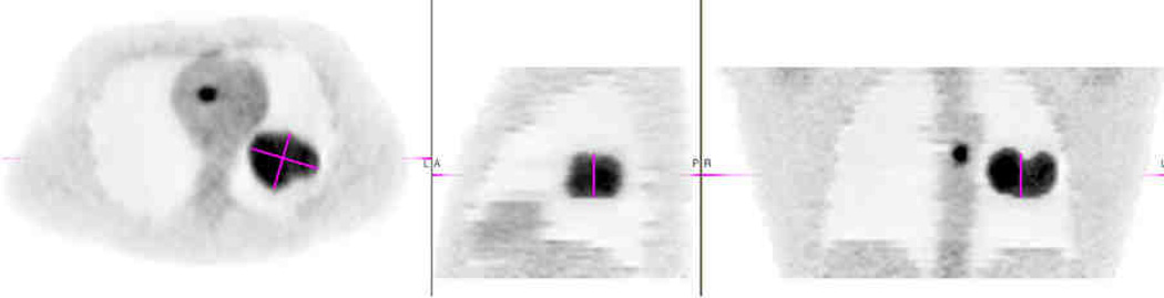
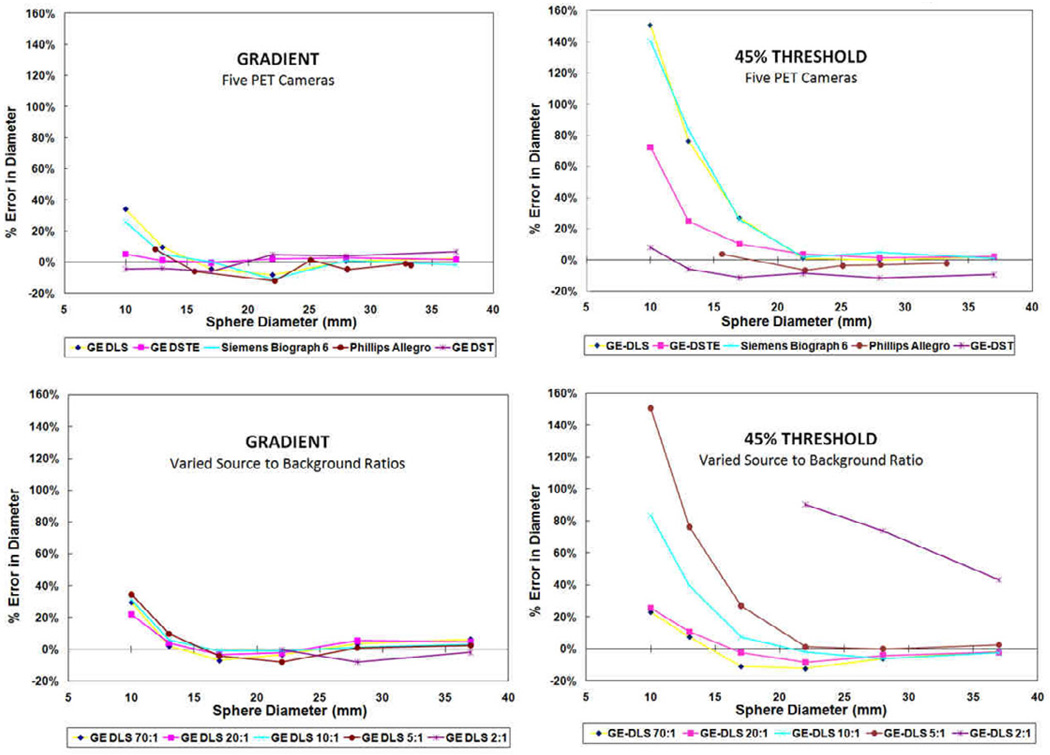
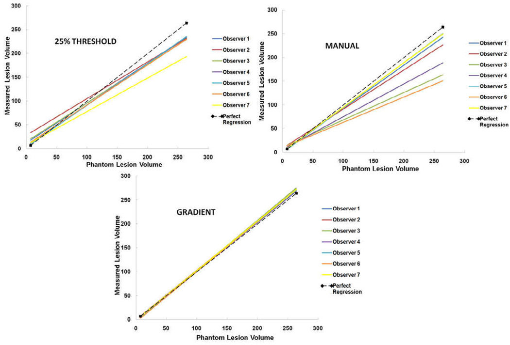
Methods and materials: Contouring accuracy was evaluated with sphere phantoms and clinically realistic Monte Carlo PET phantoms of the thorax. The sphere phantoms were 10-37 mm in diameter and were acquired at five institutions emulating clinical conditions. One institution also acquired a sphere phantom with multiple source-to-background ratios of 2:1, 5:1, 10:1, 20:1, and 70:1. One observer segmented (contoured) each sphere with GRADIENT and THRESHOLD from 25% to 50% at 5% increments. Subsequently, seven physicians segmented 31 lesions (7-264 mL) from 25 digital thorax phantoms using GRADIENT, THRESHOLD, and MANUAL.
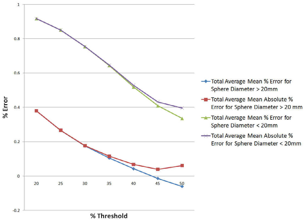
Results: For spheres <20 mm in diameter, GRADIENT was the most accurate with a mean absolute % error in diameter of 8.15% (10.2% SD) compared with 49.2% (51.1% SD) for 45% THRESHOLD (p < 0.005). For larger spheres, the methods were statistically equivalent. For varying source-to-background ratios, GRADIENT was the most accurate for spheres >20 mm (p < 0.065) and <20 mm (p < 0.015). For digital thorax phantoms, GRADIENT was the most accurate (p < 0.01), with a mean absolute % error in volume of 10.99% (11.9% SD), followed by 25% THRESHOLD at 17.5% (29.4% SD), and MANUAL at 19.5% (17.2% SD). GRADIENT had the least systematic bias, with a mean % error in volume of -0.05% (16.2% SD) compared with 25% THRESHOLD at -2.1% (34.2% SD) and MANUAL at -16.3% (20.2% SD; p value <0.01). Interobserver variability was reduced using GRADIENT compared with both 25% THRESHOLD and MANUAL (p value <0.01, Levene's test).
Conclusion: GRADIENT was the most accurate and consistent technique for target volume contouring. GRADIENT was also the most robust for varying imaging conditions. GRADIENT has the potential to play an important role for tumor delineation in radiation therapy planning and response assessment.
Copyright © 2012. Published by Elsevier Inc.
Conflict of interest statement
Conflicts of Interest Notification
Figures
Comment in
-
Gradient-PET based delineation may be improved with combined post contrast high resolution CT scan: in regard to Werner-Wasik M et al. (Int J Radiat Oncol Biol Phys 2011 Apr 28).Int J Radiat Oncol Biol Phys. 2012 Jan 1;82(1):496; author reply 496-7. doi: 10.1016/j.ijrobp.2011.06.2005. Int J Radiat Oncol Biol Phys. 2012. PMID: 22182725 No abstract available.
References
-
- Black QC, Grills IS, Kestin LL, et al. Defining a radiotherapy target with positron emission tomography. International journal of radiation oncology, biology, physics. 2004;60(4):1272–1282. - PubMed
-
- Bradley J, Thorstad WL, Mutic S, et al. Impact of FDG-PET on radiation therapy volume delineation in non–small-cell lung cancer. International journal of radiation oncology, biology, physics. 2004;59(1):78–86. - PubMed
-
- Shankar LK, Hoffman JM, Bacharach S, et al. Consensus recommendations for the use of 18F-FDG PET as an indicator of therapeutic response in patients in National Cancer Institute Trials. J Nucl Med. 2006;47(6):1059–1066. - PubMed
-
- Hoekstra CJ, Paglianiti I, Hoekstra OS, et al. Monitoring Response to Therapy in Cancer Using [18F]-2-fluoro-2-deoxy-D-glucose and Positron Emission Tomography: an Overview of Defferent Analytical Methods. European Journal of Nuclear Medicine and Molecular Imaging. 2000;27(6):731–743. - PubMed
-
- Caldwell CB, Mah K, Ung YC, et al. Observer variation in contouring gross tumor volume in patients with poorly defined non-small-cell lung tumors on CT: the impact of 18FDG-hybrid PET fusion. International journal of radiation oncology, biology, physics. 2001;51(4):923–931. - PubMed
Publication types
MeSH terms
Grants and funding
LinkOut - more resources
Full Text Sources
Medical
