. 2011 Jun;29(7):965-73.
doi: 10.1016/j.jmgm.2011.04.001. Epub 2011 Apr 13.

Novel ligands that target the mitochondrial membrane protein mitoNEET

Affiliations

Novel ligands that target the mitochondrial membrane protein mitoNEET

Robert M Bieganski et al. J Mol Graph Model. 2011 Jun.

Abstract

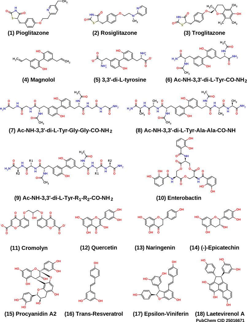
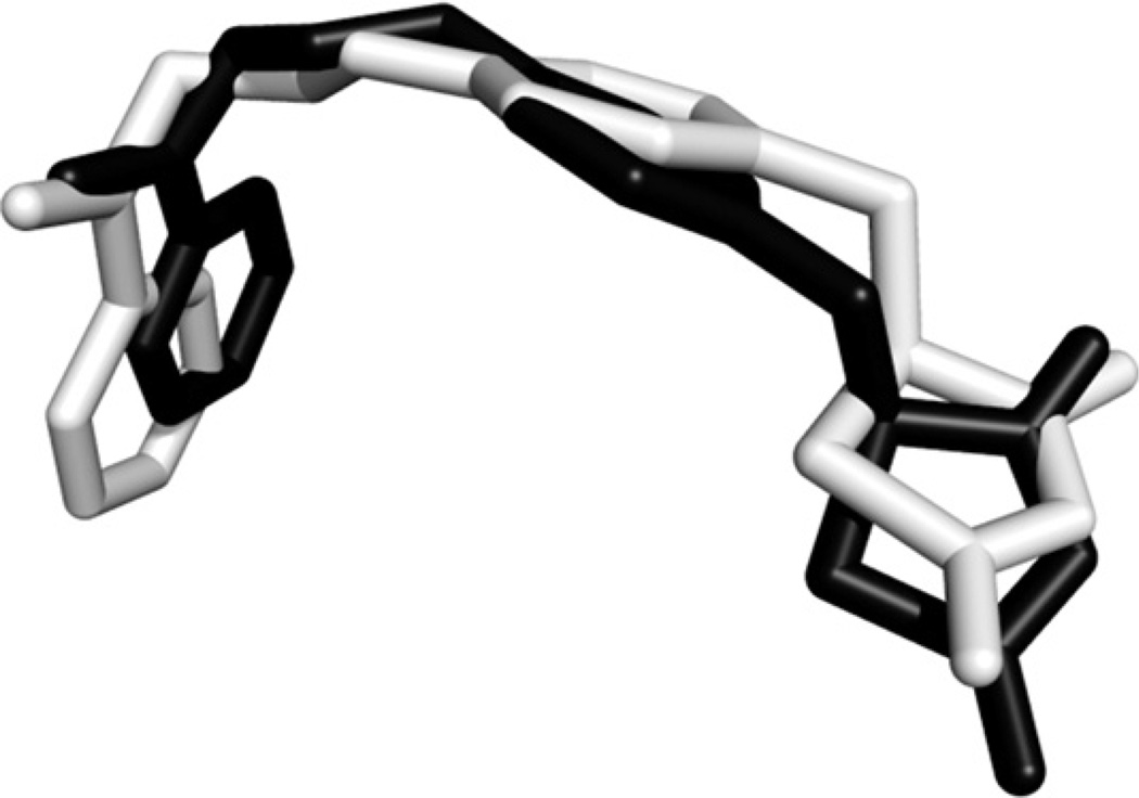
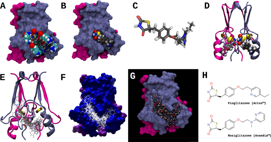
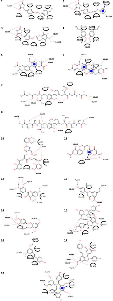
Ligands of the thiazolidinedione (TZD) class of compounds, pioglitazone (Actos™) and rosiglitazone (Avandia™) are currently approved for treatment of type 2 diabetes and are known to bind to the PPAR-γ nuclear receptor subtype. Recent evidence suggesting PPAR-γ independent action of the TZDs led to the discovery of a novel integral outer mitochondrial membrane protein, mitoNEET. In spite of the several reported X-ray crystal structures of the unbound form of mitoNEET, the location and nature of the mitoNEET ligand binding sites (LBS) remain unknown. In this study, a molecular blind docking (BD) method was used to discover potential mitoNEET LBS and novel ligands, utilizing the program AutoDock Vina (v 1.0.2). Validation of BD was performed on the PPAR-γ receptor (PDB ID: 1ZGY) with the test compound rosiglitazone, demonstrating that the binding conformation of rosiglitazone determined by AutoDock Vina matches well with that of the cocrystallized ligand (root mean square deviation of the heavy atoms 1.45Å). The locations and a general ligand binding interaction model for the LBS were determined, leading to the discovery of novel mitoNEET ligands. An in vitro fluorescence binding assay utilizing purified recombinant mitoNEET protein was used to determine the binding affinity of a predicted mitoNEET ligand, and the data obtained is in good agreement with AutoDock Vina results. The discovery of potential mitoNEET ligand binding sites and novel ligands, opens up the possibility for detailed structural studies of mitoNEET-ligand complexes, as well as rational design of novel ligands specifically targeted for mitoNEET.


Figures

Fig. 1
Fig. 1
Structures of 15 compounds predicted to bind to mitoNEET, as well as the structures of pioglitazone (1) and rosiglitazone (2).
Fig. 2
Fig. 2
Superposition of the computed rosiglitazone binding conformation (black) docked in the PPAR-γ receptor ligand binding pocket, and the cocrystallized ligand (light gray). RMSD calculation was based on the heavy atoms, and was performed using the “rmsd” command of UCSF Chimera (http://www.cgl.ucsf.edu/chimera). PPAR-γ receptor PDB ID: 1ZGY.
Fig. 3
Fig. 3
(A) Molecular surface representation of the MitoNEET homodimer; residues that interact with both pioglitazone and rosiglitazone are shown in space-filling representation, and include His48, Ile49, Gln50, Arg76, Lys78, Ala86, Lys89, and His90. Both pioglitazone and rosiglitazone are shown as licorice models, docked in the predicted binding conformation found using AutoDock Vina. Protomer I of the mitoNEET homodimer, is colored light blue, and protomer II is colored magenta. (B) Surface representation of MitoNEET homodimer, showing rosiglitazone docked (space-filling representation) in the MitoNEET binding pocket. (C) Superposition of pioglitazone and rosiglitazone in their docked conformations, as in (A); note that conformations of the TZD ring is nearly identical for both molecules. (D) Ribbon representation of the MitoNEET-rosiglitazone complex, showing rosiglitazone docked to both MitoNEET protomers; the 2Fe–2S clusters are shown in balls and sticks, where iron and sulfur atoms are colored brown and yellow, respectively. (E–G) Ribbon and surface representations of MitoNEET and an ensemble of 17 docked ligands (interactions shown in Fig. 5). (H) The structures of Pioglitazone (Actos™) and Rosiglitazone (Avandia™). Pioglitazone was the first ligand shown to bind to MitoNEET [1].
Fig. 4
Fig. 4
Schematic representations of hydrogen bonding, van der Waals (vdW), and aromatic ring interactions for the ligands listed in Fig. 1, arranged in the same numbering order. Dotted lines are hydrogen bonds, circled residues indicate van der Waals contacts with the ligands, and circles represent aromatic ring interactions (the line indicates the residue interacting with the ligand).
Fig. 5
Fig. 5
Graphical illustration depicting the relationship of the o,o′-biphenol and chromen-4-one core structures. “R” represents any functional group.
Fig. 6
Fig. 6
Fluorescence emission spectra of 2.4 × 10⁻⁵ mol/L mitoNEET in the presence of various concentrations of magnolol (T = 298 K; λex = 295 nm); a–o: [magnolol] × 10−6 mol/L = 0; 1.9; 3.7; 5.5; 7.4; 10.9; 14.4; 21.2; 27.8; 34.1; 40.2; 46.1; 57.2; 67.6; 77.4.
Fig. 7
Fig. 7
Magnolol bound to the mitoNEET receptor protein. The two iron–sulfur clusters are shown as ball and stick, while the two Trp75 residues and magnolol are shown in licorice. The intrinsic fluorescence of the two Trp75 residues, and their proximity to the binding pocket, enables fluorescence quenching.
Fig. 8
Fig. 8
Modified Stern–Volmer plot of F0/(F0F) versus 1/[Q], generated from fluorescence data in Fig. 6. R2 = 0.995.

References

    1. Colca JR, McDonald WG, Waldon DJ, Leone JW, Lull JM, Bannow CA, Lund ET, Mathews WR. Identification of a novel mitochondrial protein (”mitoNEET“) cross-linked specifically by a thiazolidinedione photoprobe. Am. J. Physiol. Endocrinol. Metab. 2004 Feb 2;286:E252–E260. - PubMed
    1. Paddock ML, Wiley SE, Axelrod HL, Cohen AE, Roy M, Abresch EC, Capraro D, Murphy AN, Nechushtai R, Dixon JE, Jennings PA. MitoNEET is a uniquely folded 2Fe 2S outer mitochondrial membrane protein stabilized by pioglitazone. Proc. Natl. Acad. Sci. U.S.A. 2007;104:14342–14347. - PMC - PubMed
    1. Bak DW, Zuris JA, Paddock ML, Jennings PA, Elliott SJ. Redox characterization of the FeS protein MitoNEET and impact of thiazolidinedione drug binding. Biochemistry. 2009;48:10193–10195. (November 43) - PMC - PubMed
    1. Wiley SE, Murphy AN, Ross SA, van der Geer P, Dixon JE. MitoNEET is an iron-containing outer mitochondrial membrane protein that regulates oxidative capacity. Proc. Natl. Acad. Sci. U.S.A. 2007 Mar 13;104:5318–5323. - PMC - PubMed
    1. Brunmair B, Staniek K, Gras F, Scharf N, Althaym A, Clara R, Roden M, Gnaiger E, Nohl H, Waldhäusl W, Fürnsinn C. Thiazolidinediones, like metformin, inhibit respiratory complex I: a common mechanism contributing to their antidiabetic actions? Diabetes. 2004 Apr 4;53:1052–1059. - PubMed

MeSH terms

LinkOut - more resources