

Structural neuroanatomy of tinnitus and hyperacusis in semantic dementia
- PMID: 21531705
- PMCID: PMC3188784
- DOI: 10.1136/jnnp.2010.235473
Structural neuroanatomy of tinnitus and hyperacusis in semantic dementia
Abstract
Introduction: Tinnitus and hyperacusis are common symptoms of excessive auditory perception in the general population; however, their anatomical substrates and disease associations continue to be defined.
Patients: with semantic dementia (SemD) frequently report tinnitus and hyperacusis but the significance and basis for these symptoms have not been elucidated.
Methods: 43 patients with a diagnosis of SemD attending a specialist cognitive disorders clinic were retrospectively studied. 14 patients (32% of the cohort) reported at least moderately severe chronic auditory symptoms: seven had tinnitus and a further seven had hyperacusis, and all had brain MRI while symptomatic. MRI data from SemD patients with and without auditory symptoms were compared using voxel based morphometry in order to identify neuroanatomical associations of tinnitus and hyperacusis.
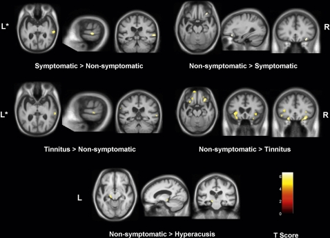
Results: Compared with SemD patients with no history of auditory symptoms, patients with tinnitus or hyperacusis had relative preservation of grey matter in the posterior superior temporal lobe and reduced grey matter in the orbitofrontal cortex and medial geniculate nucleus.
Conclusions: Tinnitus and hyperacusis may be a significant issue in SemD. Neuroanatomical evidence in SemD supports previous work implicating a distributed cortico-subcortical auditory and limbic network in the pathogenesis of these abnormal auditory percepts.
Conflict of interest statement
Figures
References
-
- Andersson G, Lindvall N, Hursti T, et al. Hypersensitivity to sound (hyperacusis): a prevalence study conducted via the Internet and post. Int J Audiol 2002;41:545–54 - PubMed
-
- Davis AC. The prevalence of hearing impairment and reported hearing disability among adults in Great Britain. Int J Epidemiol 1989;18:911–17 - PubMed
-
- Demeester K, van Wieringen A, Hendrickx JJ, et al. Prevalence of tinnitus and audiometric shape. B-ENT 2007;(Suppl 7):37–49 - PubMed
-
- Newman CW, Jacobson GP, Spitzer JB. Development of the Tinnitus Handicap Inventory. Arch Otolaryngol Head Neck Surg 1996;122:143–8 - PubMed
-
- Eggermont J, Roberts L. The neuroscience of tinnitus. Trends Neurosci 2004;27:676–82 - PubMed
Publication types
MeSH terms
Grants and funding
LinkOut - more resources
Full Text Sources
Medical