. 2011 Jul 1;286(26):23102-12.
doi: 10.1074/jbc.M110.212662. Epub 2011 Apr 29.

Sprouty-related Ena/vasodilator-stimulated phosphoprotein homology 1-domain-containing protein (SPRED1), a tyrosine-protein phosphatase non-receptor type 11 (SHP2) substrate in the Ras/extracellular signal-regulated kinase (ERK) pathway

Affiliations

Sprouty-related Ena/vasodilator-stimulated phosphoprotein homology 1-domain-containing protein (SPRED1), a tyrosine-protein phosphatase non-receptor type 11 (SHP2) substrate in the Ras/extracellular signal-regulated kinase (ERK) pathway

Martina Quintanar-Audelo et al. J Biol Chem. .

Abstract

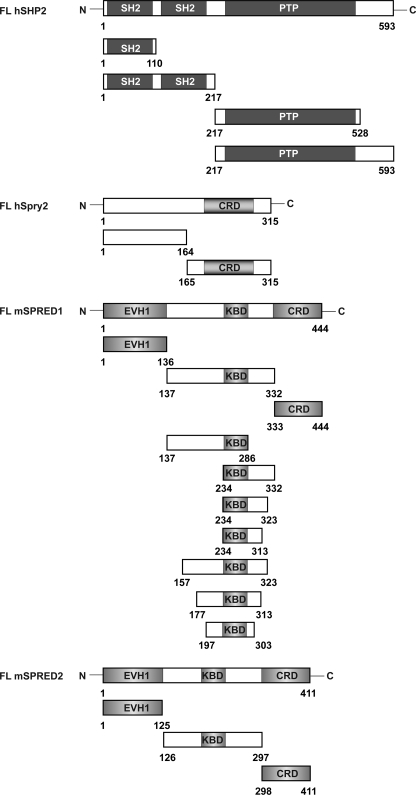
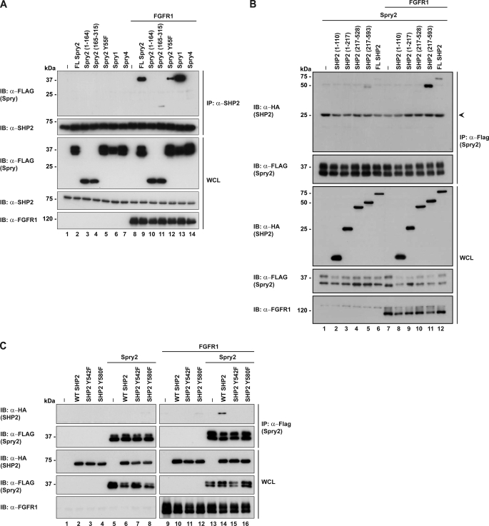
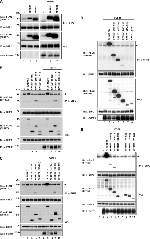
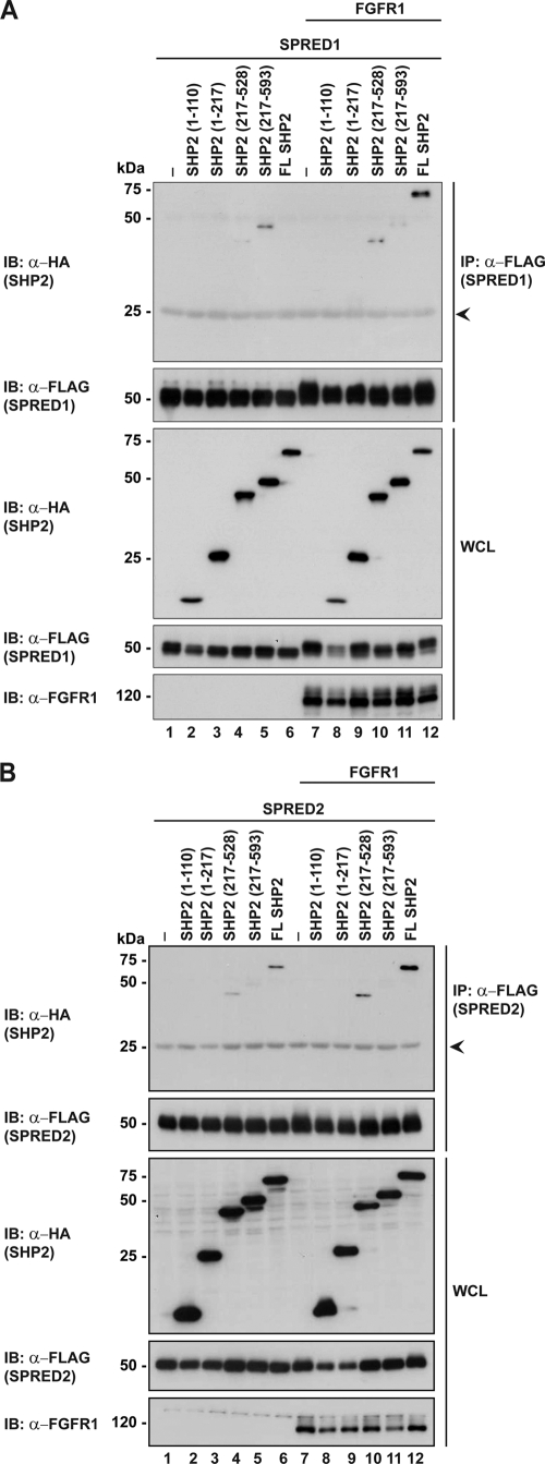
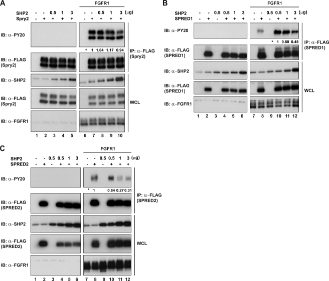
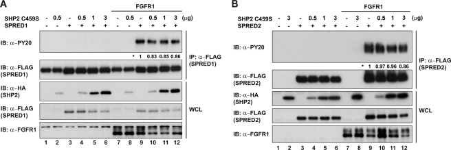
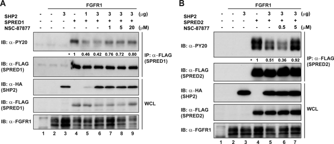
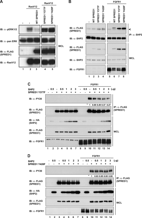
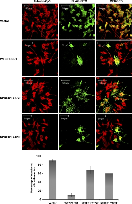
SHP2 is a tyrosine phosphatase involved in the activation of the Ras/ERK signaling pathway downstream of a number of receptor tyrosine kinases. One of the proposed mechanisms involving SHP2 in this context is to dephosphorylate and inactivate inhibitors of the Ras/ERK pathway. Two protein families bearing a unique, common domain, Sprouty and SPRED proteins, are possible candidates because they have been reported to inhibit the Ras/ERK pathway upon FGF activation. We tested whether any of these proteins are likely substrates of SHP2. Our findings indicate that Sprouty2 binds to the C-terminal tail of SHP2, which is an unlikely substrate binding site, whereas SPRED proteins bind to the tyrosine phosphatase domain that is known to be the binding site for its substrates. Overexpressed SHP2 was able to dephosphorylate SPREDs but not Sprouty2. Finally, we found two tyrosine residues on SPRED1 that are required, when phosphorylated, to inhibit Ras/ERK activation and identified Tyr-420 as a specific dephosphorylation target of SHP2. The evidence obtained indicates that SPRED1 is a likely substrate of SHP2, whose tyrosine dephosphorylation is required to attenuate the inhibitory action of SPRED1 in the Ras/ERK pathway.


Figures

FIGURE 1.
FIGURE 1.
Schematic representation of the domain structures of SHP2, Spry2, SPRED1, and SPRED2. FL hSHP2, full-length human SHP2, SH2, and PTP. FL hSpry2, full-length human Spry2, CRD. FL mSPRED1, full-length mouse SPRED1 and SPRED2, Ena-vasodilator-stimulated phosphoprotein homology-1 domain (EVH1), KBD, and CRD. These constructs were used in the subsequent figures.
FIGURE 2.
FIGURE 2.
Spry2 binds to the C-terminal tail of SHP2. A, HEK293 cells were transfected with the indicated plasmids (FLAG-Sprys, FGFR1) or the pXJ40 vector control. 24 h post-transfection, cell lysates were subjected to immunoprecipitation (IP) with anti-SHP2. Immunoprecipitates were separated in SDS-PAGE and immunoblotted (IB) with the antibodies indicated on the left. Whole cell lysates (WCL) were immunoblotted to verify equal protein expression levels. B, deletion mutants of SHP2 were co-expressed with Spry2 and FGFR1 on lanes 7–12. Lysates were immunoprecipitated with anti-FLAG, and blots were tested with the antibodies indicated on the left. C, FLAG-Spry2 and HA-SHP2 mutants were co-expressed in HEK293 cells. Lysates were immunoprecipitated with anti-FLAG and immunoblotted with anti-HA to probe for interactions with SHP2. Arrow indicates the immunoglobulin light chain.
FIGURE 3.
FIGURE 3.
SPRED1 and SPRED2 interact with SHP2 through via amino acids 135–157. A, endogenous SHP2 was immunoprecipitated in cells overexpressing FLAG-tagged SPRED1 and SPRED2 with or without FGFR1 overexpressed. Lysates were processed as described in Fig. 2A. Immunoblots were incubated with anti-FLAG to prove for interactions with SHP2. B and C, FLAG-SPRED1 and FLAG-SPRED2, respectively, full-length or deletion mutants were transfected in HEK293 cells, and endogenous SHP2 was immunoprecipitated with anti-SHP2. Immunoprecipitates were separated in SDS-PAGE. D and E, HEK293 lysates expressing SPRED1 mutants as indicated on the panels were immunoprecipitated with anti-SHP2 to pull down endogenous SHP2. Immunoprecipitates were separated in SDS-PAGE and probed with anti-FLAG to detect binding. Arrowheads indicate the immunoglobulin heavy chain. IP, immunoprecipitation; IB, immunoblot; WCL, whole cell lysate.
FIGURE 4.
FIGURE 4.
SPRED1 and SPRED2 bind to the PTP domain of SHP2. A, FLAG-SPRED1 was co-expressed with HA-SHP2 full-length or deletion mutants. Lysates were immunoprecipitated with anti-FLAG and evaluated by Western blotting. B, FLAG-SPRED2 was co-expressed together with HA-SHP2 full-length or deletion mutants, and samples were immunoprecipitated with anti-FLAG and analyzed by Western blotting with the indicated antibodies on the left. Arrowheads indicate the immunoglobulin light chain. IP, immunoprecipitation; IB, immunoblot; WCL, whole cell lysate.
FIGURE 5.
FIGURE 5.
SHP2 dephosphorylates SPRED1 and SPRED2 but not Spry2. A, HEK293 cells were transfected with FLAG-Spry2 and increasing concentrations of SHP2 with or without FGFR1 activation. Lysates were immunoprecipitated with anti-FLAG, and immunoblots were probed with anti-PY20 to detect tyrosine phosphorylation levels. B, cell lysates co-transfected with FLAG-SPRED1 and SHP2 were treated as mentioned in A. C, cell lysates co-transfected with FLAG-SPRED2 and SHP2 were treated as mentioned in A. Numbers next to the asterisk indicate quantification results of tyrosine phosphorylation levels on the PY20 blot. SHP2 band on the anti-SHP2 blots (A–C) on lanes 1 and 2 represent endogenous SHP2 detected by the antibody. IP, immunoprecipitation; IB, immunoblot; WCL, whole cell lysate.
FIGURE 6.
FIGURE 6.
SHP2C459S does not dephosphorylate SPRED. A, lysates co-expressing FLAG-SPRED1 and increasing concentrations of HA-SHP2C459S were immunoprecipitated with anti-FLAG. Immunoblots were probed with anti-PY20 to detect tyrosine phosphorylation levels. B, cells co-expressing FLAG-SPRED2 and HA-SHP2C459S were treated as mentioned in A. Numbers next to the asterisk indicate quantification of the tyrosine phosphorylation levels on the PY20 blot. IP, immunoprecipitation; IB, immunoblot; WCL, whole cell lysate.
FIGURE 7.
FIGURE 7.
SHP2 phosphatase inhibition rescues tyrosine phosphorylation of SPRED. A, HEK293 cells were transfected with FLAG-SPRED1 and HA-SHP2. 24 h post-transfection, cells were treated with SHP2 inhibitor NSC-87877 for 2 h (lanes 7–9). Lysates were immunoprecipitated with anti-FLAG, subjected to SDS-PAGE, and immunoblotted as indicated. B, FLAG-SPRED2 and HA-SHP2 overexpressed in HEK293 cells were treated with NSC-87877 (lanes 5–7) for 2 h before harvesting. Lysates were treated as mentioned in A. Numbers next to the asterisk indicate quantification of tyrosine phosphorylation levels on the PY20 blot. IP, immunoprecipitation; IB, immunoblot; WCL, whole cell lysate.
FIGURE 8.
FIGURE 8.
SHP2 dephosphorylates Tyr-420 on SPRED1. A, HEK293 cells were transfected with the indicated plasmids (FLAG-SPRED and RasV12) or the pXJ40 vector control. Whole cell lysates (WCL) were analyzed by Western blotting with the indicated antibodies on the left. B, endogenous SHP2 was immunoprecipitated, with anti-SHP2, in cells overexpressing FLAG-SPRED1 WT or respective mutants. Immunoprecipitates were resolved by SDS-PAGE. C, FLAG-SPRED1Y377F was co-transfected with increasing concentrations of HA-SHP2 (0.5–3 μg), and lysates were immunoprecipitated with anti-FLAG. Immunoblots were probed with the antibodies indicated on the left. D, cells overexpressing FLAG-SPRED1Y420F and HA-SHP2 were treated as mentioned in A. Numbers next to the asterisk indicate quantification of the tyrosine phosphorylation levels on the PY20 blot. The arrowhead indicates the light band at 50 kDa corresponding to the immunoglobulin heavy chain.
FIGURE 9.
FIGURE 9.
Tyrosines 377 and 420 of SPRED1 are required for inhibition of basic fibroblast growth factor-induced neurite outgrowth in PC12 cells. PC12 cells grown on poly-l-lysine-coated coverslips were transfected with FLAG-SPRED1, FLAG-SPRED1Y377F, or FLAG-SPRED1Y420F, after transfection cells were stimulated with basic fibroblast growth factor as described in “Experimental Procedures.” The SPRED1 protein was stained with FLAG-FITC, and the cells were counterstained with Cy3-conjugated anti-tubulin. Scale bar, 50 μm. The average percentage of transfected cells bearing neurites from a minimum of three independent experiments is graphically shown in the bottom panels.

Similar articles

Cited by

References

    1. Schlessinger J. (2002) Cell 110, 669–672 - PubMed
    1. Malumbres M., Barbacid M. (2003) Nat. Rev. Cancer 3, 459–465 - PubMed
    1. Eswarakumar V. P., Lax I., Schlessinger J. (2005) Cytokine Growth Factor Rev. 16, 139–149 - PubMed
    1. Hadari Y. R., Kouhara H., Lax I., Schlessinger J. (1998) Mol. Cell. Biol. 18, 3966–3973 - PMC - PubMed
    1. Araki T., Nawa H., Neel B. G. (2003) J. Biol. Chem. 278, 41677–41684 - PubMed

Publication types

MeSH terms

Substances

LinkOut - more resources