. 2011 Jun 17;286(24):21555-64.
doi: 10.1074/jbc.M111.224048. Epub 2011 Apr 29.

A20 is induced by Kaposi sarcoma-associated herpesvirus-encoded viral FLICE inhibitory protein (vFLIP) K13 and blocks K13-induced nuclear factor-kappaB in a negative feedback manner

Affiliations

A20 is induced by Kaposi sarcoma-associated herpesvirus-encoded viral FLICE inhibitory protein (vFLIP) K13 and blocks K13-induced nuclear factor-kappaB in a negative feedback manner

Hittu Matta et al. J Biol Chem. .

Abstract

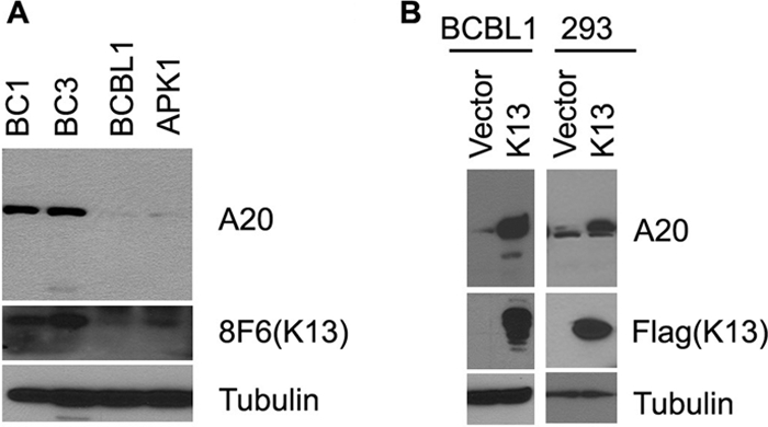
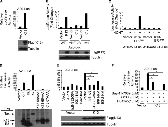
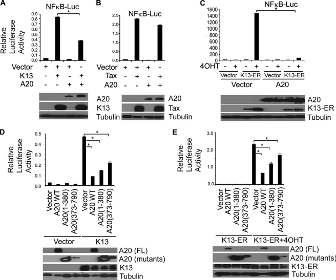
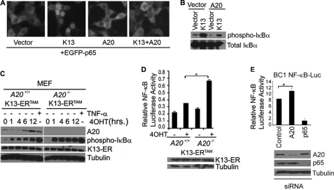
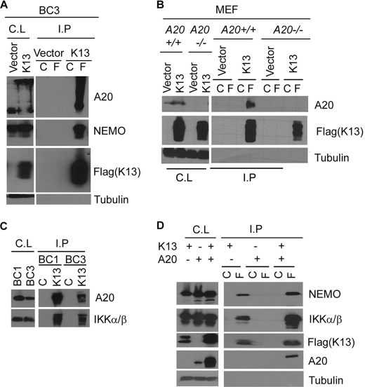
Expression of A20, a negative regulator of the NF-κB pathway, is frequently lost in several subtypes of Hodgkin and non-Hodgkin lymphoma. We report that A20 is expressed in Kaposi sarcoma-associated herpesvirus (KSHV)-infected primary effusion lymphoma cell lines, and its expression correlates closely with the expression of KSHV-encoded viral FLICE inhibitory protein K13. Ectopic expression of K13 induced A20 expression through NF-κB-mediated activation of A20 promoter. In turn, A20 blocked K13-induced NF-κB activity and up-regulation of proinflammatory cytokines CCL20 and IL-8 in a negative feedback fashion. Both the N-terminal deubiquitinating domain and the C-terminal zinc finger domain of A20 were involved in the inhibition of K13-induced NF-κB activity. Overexpression of A20 blocked K13-induced IκBα phosphorylation, NF-κB nuclear translocation, and cellular transformation. Consistent with the above, K13-induced IκBα phosphorylation and NF-κB transcriptional activation were enhanced in A20-deficient cells. Finally, A20 was found to interact physically with K13. Taken collectively, these results demonstrate that K13 is a key determinant of A20 expression in KSHV-infected cells, and A20 is a key negative regulator of K13-induced NF-κB activity. A20 might serve to control the inflammatory response to KSHV infection and protect KSHV-infected cells from apoptosis.


Figures

FIGURE 1.
FIGURE 1.
K13 up-regulates A20 expression in PEL cell lines. A, Western blots showing expression of A20 and K13 in PEL cell lines. B, Western blots showing up-regulation of A20 in BCBL1 and 293 cells by ectopic K13 expression. Stable pools of BCBL1 and 293 cells expressing an empty vector and FLAG-K13 were generated by retroviral gene transfer. Cell lysates from these cells were used for Western blot analysis. Tubulin blot shows equal protein loading (bottom panel).
FIGURE 2.
FIGURE 2.
K13 activates A20 promote through NF-κB pathway. A and B, the 293T cells were transfected with a control vector and a vector encoding FLAG-K13 (250 ng/ml) along with either A20-Luc or A20 mNF-κB-Luc or A20 m1-Luc reporter construct (75 ng/well) and a pRSV/LacZ (β-galactosidase) reporter construct (75 ng/well), and the reporter assay was performed as described under “Materials and Methods.” The values shown are averages (mean ± S.E.) of one representative experiment of three in which each transfection was performed in duplicate. Expression of the K13 was confirmed by Western blot analysis with an antibody against the FLAG epitope tag. C, K13-ERTAM induces A20 promoter activity upon treatment with 4OHT. The experiment was performed essentially as described in A. The values shown are averages (mean ± S.E.) of one representative experiment ofthree in which each transfection was performed in duplicate. D, wild-type K13 but not vFLIP E8- or NF-κB-defective mutants of K13 induces A20 promoter activity. 293T cells were transfected with the indicated FLAG-tagged expression constructs along with a A20 promoter luciferase construct. The experiment was performed as described for A. The values shown are averages (mean ± S.E.) of one representative experiment of three in which each transfection was performed in duplicate. Lower panel shows the expression of the transfected proteins as dermined by Western blotting with a FLAG antibody. E, dominant negative mutants of IκBα (IκBαΔN and IκBαS32A/S36A), IKK1 and IKK2 block K13-induced A20 promoter activity. The 293T cells were transfected either with an empty vector or K13, along with an A20 luciferase reporter construct and a reporter construct, as described in A. The amount of inhibitor plasmids (500 ng/well) was five times the amount of vector or K13 (100 ng/well) plasmid, and the total amount of transfected DNA was kept constant by adding empty vector. The values shown are averages (mean ± S.E.) of one representative experiment of three in which each transfection was performed in duplicate. Expression of the K13 was confirmed by Western blot analysis with the FLAG antibody. Asterisks indicate significance at levels of p ≤ 0.05. F, pharmacologic inhibitors of NF-κB block K13-induced A20 promoter activation. The 293T cells were transfected with an empty vector or a vector encoding K13 along with A20-Luc and pRSV/LacZ reporter constructs. Approximately 3 h after transfection, cells were treated with dimethyl sulfoxide (vehicle) or the indicated compounds for 18 h before cell lysis and measurement of reporter activities. Asterisks indicate significance at levels of p ≤ 0.05 compared with vehicle-treated cells.
FIGURE 3.
FIGURE 3.
A20 inhibits K13-induced NF-κB activity. A and B, 293T cells were transfected with control vector, K13, or Tax along with an NF-κB-Luc construct (75 ng/well) and a pRSV/LacZ (β-galactosidase) reporter construct (75 ng/well) in the presence or absence of A20. Cells were lysed 20 h after transfection for reporter assays. The values shown are the averages (mean ± S.E.) of one representative experiment of three in which each transfection was performed in duplicate. The expression of the transfected A20 and FLAG-tagged K13 and Tax proteins was demonstrated by immunoblotting (lower panel). C, A20 inhibits K13-ERTAM-induced NF-κB activity. The experiment was performed essentially as described in 2A. The values shown are averages (mean ± S.E.) of one representative experiment of three in which each transfection was performed in duplicate. D and E, 293T cells were transfected with K13 or K13-ERTAM along with NF-κB-Luc and pRSV/LacZ reporter constructs in the presence or absence of the indicated A20 constructs. The cells transfected with K13-ERTAM were treated with 4OHT (20 nm) for 24 h prior to cell lysis and measurement of reporter activities. The values shown are the averages (mean ± S.E.) of one representative experiment of three in which each transfection was performed in duplicate. Lower panel shows the expression of the transfected proteins as determined by Western blotting with a FLAG antibody. Asterisks indicate statistical significance at levels of p ≤ 0.05.
FIGURE 4.
FIGURE 4.
A20 blocks K13-induced up-regulation of CCL20 and IL-8 and cellular transformation. A–D, A20 blocks K13-induced up-regulation of CCL20 and IL-8. 293T cells were transfected with an empty vector, K13, or K13-ERTAM along with luciferase reporter constructs driven by CCL20 (CCL20-Luc) or IL-8 promoters (IL-8-Luc) (75 ng/well) and a pRSV/LacZ (β-galactosidase) reporter construct (75 ng/well) in the presence or absence of A20. The cells transfected with K13-ERTAM were subsequently treated with 4OHT (20 nm) for 24 h and the luciferase reporter assay performed essentially as described in Fig. 2A. Lower panels demonstrate the expression of the transfected proteins as measured by immunoblotting with the indicated antibodies. The values shown are the averages (mean ± S.E.) of one representative experiment of three in which each transfection was performed in duplicate. E and F, cellular supernatants from 293T cells transfected, as explained in A–D, were collected before doing luciferase assay and used to measure the secretion of CCL20 and IL-8 by ELISA. The values shown are averages (mean ± S.E.) of one representative experiment of three in which the level of CCL20 or IL-8 secretion was measured in duplicate. Asterisks (*) indicate significance at levels of p ≤ 0.05. G, immunoblot shows increased A20 expression in Rat1-K13 cells transfected with an A20 expression construct. H, soft agar colony formation by Rat1-K13 cells transfected with an A20 expression vector was reduced compared with those transfected with an empty vector. The values shown are mean ± S.D. of two independent experiments performed in triplicate. *, p ≤ 0.05 versus vector cells.
FIGURE 5.
FIGURE 5.
A20 blocks K13-induced NF-κB activation by blocking IκBα phosphorylation and p65 nuclear translocation. A, 293T cells were transfected with an EGFP-p65 fusion construct along with the indicated expression constructs, and cellular localization of EGFP-p65 was examined by fluorescence microscopy. K13-induced nuclear accumulation of EGFP-p65 is blocked by co-expression of A20. B, 293T cells were transfected with K13 in the presence or absence of A20, and 24 h after transfection, total cell lysates were prepared and used to examine the effect of A20 on K13-induced phosphorylation of IκBα. The expression of total IκBα is shown in the lower panel. C, immunoblot shows increase in phosphorylation of IκBα in K13-ERTAM-expressing A20−/− MEFs compared with the corresponding A20+/+ MEFs. A20+/+ and A20−/− MEFs expressing FLAG-K13-ERTAM were treated with 4OHT for the indicated time points, and the phosphorylation of IκBα and A20 expression was measured using the indicated antibodies. Equivalent expression of the FLAG-tagged K13-ERTAM protein and equal protein loading was confirmed by blotting with FLAG and tubulin antibodies, respectively. D, A20+/+ and A20−/− MEFs expressing FLAG-K13-ERTAM were transfected with NF-κB promoter luciferase construct (75 ng/well) and a Renilla reporter construct (75 ng/well, normalization control) using Lipofectamine-mediated transfection as described previously (33). The transfected cells were subsequently treated with 4OHT (20 nm) for 48 h, and the luciferase reporter assay was performed essentially as described in Fig. 2A. The values shown are the averages (mean ± S.E.) of one representative experiment of three in which each transfection was performed in duplicate. Asterisks (*) indicate significance at levels of p ≤ 0.05. E, upper, BC1-NFκB-Luc cells were transfected with a control siRNA (lamin A/C) or siRNAs directed against A20 or p65, respectively. Approximately 72 h after transfection, cell lysates were prepared and used for the measurement of luciferase activity. The data represent mean ± S.E. of a representative of two independent experiments with similar results. *, p ≤ 0.05 versus control cells. E, lower, immunoblot confirms specific knock-down of A20 and p65 proteins following siRNA-mediated gene silencing.
FIGURE 6.
FIGURE 6.
K13 interacts with A20. A, cell lysates (C.L.) prepared from BC3 cells expressing an empty vector or FLAG-tagged K13 were immunoprecipitated (I.P.) using control (C) or FLAG (F) beads, and the presence of interacting A20 and NEMO proteins was detected by Western blotting with the indicated antibodies. B, co-immunoprecipitation assay shows interaction of K13 with A20 in A20+/+ MEFs. The experiment was performed essentially as described for A. A tubulin blot shows equal protein loading. C, cell lysates (C.L.) prepared from BC1 and BC3 cells were immunoprecipitated using control antibody or 8F6 antibody raised against K13, and the presence of interacting A20 and IKK α/β proteins was detected by Western blotting with the indicated antibodies. D, A20 does not block the interaction between K13 and IKK complex subunits. 293T cells were transfected with an empty vector or a vector expressing FLAG-tagged K13, either alone or with A20, and a co-immunoprecipitation assay was performed essentially as described for A. A tubulin blot shows equal protein loading.

Similar articles

Cited by

References

    1. Aggarwal B. B. (2004) Cancer Cell 6, 203–208 - PubMed
    1. Dixit V. M., Green S., Sarma V., Holzman L. B., Wolf F. W., O'Rourke K., Ward P. A., Prochownik E. V., Marks R. M. (1990) J. Biol. Chem. 265, 2973–2978 - PubMed
    1. Hymowitz S. G., Wertz I. E. (2010) Nat. Rev. Cancer 10, 332–341 - PubMed
    1. Laherty C. D., Hu H. M., Opipari A. W., Wang F., Dixit V. M. (1992) J. Biol. Chem. 267, 24157–24160 - PubMed
    1. Laherty C. D., Perkins N. D., Dixit V. M. (1993) J. Biol. Chem. 268, 5032–5039 - PubMed

Publication types

MeSH terms