. 2011 Aug;301(2):L181-6.
doi: 10.1152/ajplung.00321.2010. Epub 2011 Apr 29.

Mucociliary interactions and mucus dynamics in ciliated human bronchial epithelial cell cultures

Affiliations

Mucociliary interactions and mucus dynamics in ciliated human bronchial epithelial cell cultures

Patrick R Sears et al. Am J Physiol Lung Cell Mol Physiol. 2011 Aug.

Abstract

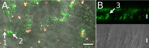
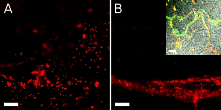
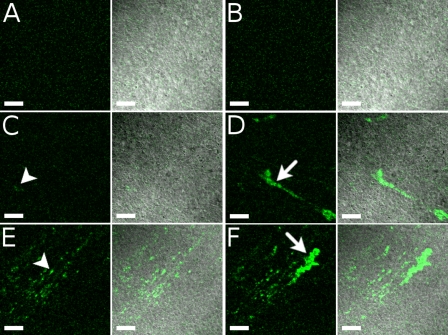
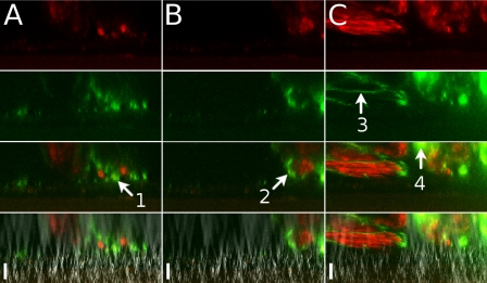
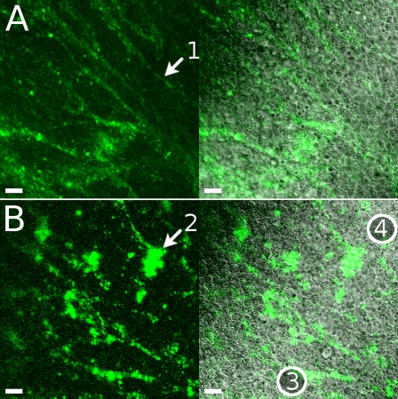
The airway epithelial surface liquid is generally considered to be composed of two layers, a periciliary layer and a continuous thick mucus layer moving in bulk. This view may not be appropriate for all areas of the lung. Our hypothesis, that mucus may form a discontinuous layer with dynamic attachments to the surface, is investigated using a culture system. We used live-cell confocal microscopy to investigate thin mucus layers and fluorescent beads and exogenous MUC5B to visualize mucus dynamics on ciliated human bronchial cultures. A continuous mucus layer was not observed. In sparsely ciliated cultures, mucus attached to ciliated cells; however, in highly ciliated cultures, mucus formed strands several hundred micrometers long. As with increases in ciliation, increases in bead concentration caused the appearance of mucus strands. We confirmed the involvement of mucins in the binding of mucus to cilia by adding labeled purified MUC5B to the cultures. These data suggest that mucins may have an intrinsic ability to form attachments to cilia. The significance of these findings is that aberrant modulation of such an intrinsic property may explain the initiation of highly adherent mucus in cystic fibrosis lung disease.


Figures

Fig. 1.
Fig. 1.
Sparsely ciliated cultures in 2 views. A: confocal x,y scan at the level of cilia. Red-fluorescent beads were 1 μm diameter; green-fluorescent beads were 20 nm diameter. B: x,z scan for fluorescence and differential interference contrast (DIC) in which only 20-nm green-fluorescent beads were present. One-micrometer beads (arrow 1) remained outside the structures labeled by 20-nm beads (arrow 2). Structures often extended for >10 μm above the cell surface (arrow 3). See Supplemental Material for 1 movie for each view (Movies 1A and 1B). Scale bars, 10 μm.
Fig. 2.
Fig. 2.
Mucus behavior on fully ciliated cultures shown as fluorescence on the culture surface from red-fluorescent 1-μm beads. A: fluorescence just after bead addition (also see Supplemental Movie 2A). B: fluorescence a few minutes later, when thick strands were moving across the field of view (also see Supplemental Movie 2B). Inset: 20-nm (green) and 1-μm (red) beads much later, when strands have stopped flowing. Scale bars, 30 μm.
Fig. 3.
Fig. 3.
Effect of bead concentration on mucus dynamics shown in 3 stages during a slow increase in concentration of 20-nm beads. Each row of panels and each movie in Supplemental Material (Supplemental Movies 3AB, 3CD, and 3EF) show a different bead concentration. Each pair of images shows fluorescence (green on black) and merged fluorescence-DIC (green on gray). Areas imaged in the 3 rows are not identical but are highly ciliated. A, C, and E: movie frame in which no fluorescence was moving across the field of view. Frame represents attached mucus. B, D, and F: frame for which the most fluorescence was moving. A and B (Supplemental Movie 3AB): culture before addition of beads. The same movie frame was used for A and B. Some autofluorescence can be seen. C and D (Supplemental Movie 3CD): culture after second addition of beads, at which point an increase in fluorescence was first seen. E and F (Supplemental Movie 3EF): culture after last addition of beads. Some attached clumps are marked with arrowheads (C and E), and some larger flowing clumps are shown with arrows (D and F). Fluorescent images of E and F were taken using a slightly lower gain. All images were processed in exactly the same way to enhance contrast. Scale bars, 40 μm.
Fig. 4.
Fig. 4.
Two-color sequential addition of beads. Red- followed by green-fluorescent 20-nm beads were added to a culture seen here in profile. From top to bottom: red fluorescence, green fluorescence, merged red and green, and merged fluorescence and DIC. At 3 min after the final wash (A), green and red fluorescence was seen in separate areas, with some green fluorescence closer to the culture surface (arrow 1). At 20 min (B), green fluorescence was often seen circumscribing areas of red fluorescence (arrow 2). At 36 min (C), some areas of fluorescence appeared thin, as if stretched (arrow 3), and other areas were starting to show combined red and green fluorescence (arrow 4). Scale bars, 23 μm.
Fig. 5.
Fig. 5.
Exogenous MUC5B. FITC-labeled exogenous MUC5B was added to a culture. Left: fluorescence; right: merged fluorescence and DIC. A: highly ciliated area a few minutes after MUC5B addition. Arrow 1, thin MUC5B strand. B: images obtained 2 h after MUC5B addition showing an area that is highly ciliated, except for a band running from bottom left (circled 3) diagonally up and to the right (circled 4). In this band, MUC5B (green) could be seen to temporarily attach to ciliated cells (gray DIC motion in Supplemental Movie 5B). Arrow 2, one of the thicker clumps that showed temporary attachment to a ciliated cell. Scale bars, 20 μm.

Similar articles

Cited by

References

    1. Armstrong DS, Hook SM, Jamsen KM, Nixon GM, Carzino R, Carlin JB, Robertson CF, Grimwood K. Lower airway inflammation in infants with cystic fibrosis detected by newborn screening. Pediatr Pulmonol 40: 500–510, 2005 - PubMed
    1. Bernacki SH, Nelson AL, Abdullah L, Sheehan JK, Harris A, Davis CW, Randell SH. Mucin gene expression during differentiation of human airway epithelia in vitro. Muc4 and Muc5b are strongly induced. Am J Respir Cell Mol Biol 20: 595–604, 1999 - PubMed
    1. Dillon RH, Fauci LJ, Omoto C, Yang X. Fluid dynamic models of flagellar and ciliary beating. Ann NY Acad Sci 1101: 494–505, 2007 - PubMed
    1. Fulcher ML, Gabriel S, Burns KA, Yankaskas JR, Randell SH. Well-differentiated human airway cultures. Methods Mol Med 107: 183–206, 2005 - PubMed
    1. Gehr P, Green FH, Geiser M, Hof VI, Lee MM, Schürch S. Airway surfactant, a primary defense barrier: mechanical and immunological aspects. J Aerosol Med 9: 163–181, 1996 - PubMed

Publication types