

Roles of cysteine proteases Cwp84 and Cwp13 in biogenesis of the cell wall of Clostridium difficile
- PMID: 21531808
- PMCID: PMC3133288
- DOI: 10.1128/JB.00248-11
Roles of cysteine proteases Cwp84 and Cwp13 in biogenesis of the cell wall of Clostridium difficile
Abstract
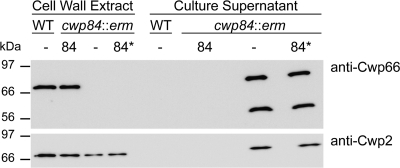
Clostridium difficile expresses a number of cell wall proteins, including the abundant high-molecular-weight and low-molecular-weight S-layer proteins (SLPs). These proteins are generated by posttranslational cleavage of the precursor SlpA by the cysteine protease Cwp84. We compared the phenotypes of C. difficile strains containing insertional mutations in either cwp84 or its paralog cwp13 and complemented with plasmids expressing wild-type or mutant forms of their genes. We show that the presence of uncleaved SlpA in the cell wall of the cwp84 mutant results in aberrant retention of other cell wall proteins at the cell surface, as demonstrated by secretion of the proteins Cwp66 and Cwp2 into the growth medium. These phenotypes are restored by complementation with a plasmid expressing wild-type Cwp84 enzyme but not with one encoding a Cys116Ala substitution in the active site. The cwp13 mutant cleaved the SlpA precursor normally and had a wild-type-like colony phenotype. Both Cwp84 and Cwp13 are produced as proenzymes which are processed by cleavage to produce mature enzymes. In the case of Cwp84, this cleavage does not appear to be autocatalytic, whereas in Cwp13 autocatalysis was demonstrated as a Cys109Ala mutant did not undergo processing. Cwp13 appears to have a role in processing of Cwp84 but is not essential for Cwp84 activity. Cwp13 cleaves SlpA in the HMW SLP domain, which we suggest may reflect a role in cleavage and degradation of misfolded proteins at the cell surface.
Figures


Similar articles
-
Characteristics of the Clostridium difficile cell envelope and its importance in therapeutics.Microb Biotechnol. 2017 Jan;10(1):76-90. doi: 10.1111/1751-7915.12372. Epub 2016 Jun 17. Microb Biotechnol. 2017. PMID: 27311697 Free PMC article. Review.
-
Cwp84, a surface-associated cysteine protease, plays a role in the maturation of the surface layer of Clostridium difficile.J Biol Chem. 2009 Dec 11;284(50):34666-73. doi: 10.1074/jbc.M109.051177. Epub 2009 Oct 6. J Biol Chem. 2009. PMID: 19808679 Free PMC article.
-
Transcription and analysis of polymorphism in a cluster of genes encoding surface-associated proteins of Clostridium difficile.J Bacteriol. 2003 Aug;185(15):4461-70. doi: 10.1128/JB.185.15.4461-4470.2003. J Bacteriol. 2003. PMID: 12867455 Free PMC article.
-
Localization of the Clostridium difficile cysteine protease Cwp84 and insights into its maturation process.J Bacteriol. 2011 Oct;193(19):5314-21. doi: 10.1128/JB.00326-11. Epub 2011 Jul 22. J Bacteriol. 2011. PMID: 21784932 Free PMC article.
-
The structure of the S-layer of Clostridium difficile.J Cell Commun Signal. 2018 Mar;12(1):319-331. doi: 10.1007/s12079-017-0429-z. Epub 2017 Nov 23. J Cell Commun Signal. 2018. PMID: 29170885 Free PMC article. Review.
Cited by
-
Clostridioides difficile infection: traversing host-pathogen interactions in the gut.Microbiology (Reading). 2023 Feb;169(2):001306. doi: 10.1099/mic.0.001306. Microbiology (Reading). 2023. PMID: 36848200 Free PMC article. Review.
-
Development of an Effective Nontoxigenic Clostridioides difficile-Based Oral Vaccine against C. difficile Infection.Microbiol Spectr. 2022 Jun 29;10(3):e0026322. doi: 10.1128/spectrum.00263-22. Epub 2022 May 18. Microbiol Spectr. 2022. PMID: 35583336 Free PMC article.
-
What's a Biofilm?-How the Choice of the Biofilm Model Impacts the Protein Inventory of Clostridioides difficile.Front Microbiol. 2021 Jun 10;12:682111. doi: 10.3389/fmicb.2021.682111. eCollection 2021. Front Microbiol. 2021. PMID: 34177868 Free PMC article.
-
Characteristics of the Clostridium difficile cell envelope and its importance in therapeutics.Microb Biotechnol. 2017 Jan;10(1):76-90. doi: 10.1111/1751-7915.12372. Epub 2016 Jun 17. Microb Biotechnol. 2017. PMID: 27311697 Free PMC article. Review.
-
Spatial organization of Clostridium difficile S-layer biogenesis.Sci Rep. 2020 Aug 24;10(1):14089. doi: 10.1038/s41598-020-71059-x. Sci Rep. 2020. PMID: 32839524 Free PMC article.
References
-
- Bartlett J. G. 2007. Clostridium difficile: old and new observations. J. Clin. Gastroenterol. 41(Suppl. 1):S24–S29
-
- Brannigan J. A., et al. 1995. A protein catalytic framework with an N-terminal nucleophile is capable of self-activation. Nature 378:416–419 - PubMed
-
- Calabi E., et al. 2001. Molecular characterization of the surface layer proteins from Clostridium difficile. Mol. Microbiol. 40:1187–1199 - PubMed
Publication types
MeSH terms
Substances
Grants and funding
LinkOut - more resources
Full Text Sources
Other Literature Sources
Molecular Biology Databases
