

Exome sequencing identifies an MYH3 mutation in a family with distal arthrogryposis type 1
- PMID: 21531865
- PMCID: PMC3102311
- DOI: 10.2106/JBJS.J.02004
Exome sequencing identifies an MYH3 mutation in a family with distal arthrogryposis type 1
Abstract
Background: Few genes responsible for distal arthrogryposis type 1 are known, although genes coding for the proteins in the sarcomere have been implicated in other types of distal arthrogryposis. Cost-effective sequencing methods are now available to examine all genes in the human genome for the purpose of establishing the genetic basis of musculoskeletal disorders.
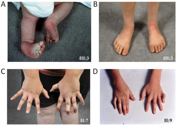
Methods: A multigenerational family with distal arthrogryposis type 1 characterized by clubfoot and mild hand contractures was identified, and exome sequencing was performed on DNA from one of the affected family members. Linkage analysis was used to confirm whether a genetic variant segregated with distal arthrogryposis.
Results: Exome sequencing identified 573 novel variants that were not present in control databases. A missense mutation in MYH3 (a gene coding for the heavy chain of myosin), causing an F437I amino acid substitution, was identified that segregated with distal arthrogryposis in this family. Linkage analysis confirmed that this MYH3 mutation was the only exome variant common to all six affected individuals.
Conclusions: Identification of an MYH3 mutation in this family with distal arthrogryposis type 1 broadens the phenotype associated with MYH3 mutations to include distal arthrogryposis types 1, 2A (Freeman-Sheldon syndrome), and 2B (Sheldon-Hall syndrome). Exome sequencing is a useful and cost-effective method to discover causative genetic mutations, although data from extended families may be needed to confirm the importance of the hundreds of identified variants.
Figures


References
-
- Bamshad M, Jorde LB, Carey JC. A revised and extended classification of the distal arthrogryposes. Am J Med Genet. 1996;65:277-81 - PubMed
-
- Hall JG. Genetic aspects of arthrogryposis. Clin Orthop Relat Res. 1985;194:44-53 - PubMed
-
- Toydemir RM, Rutherford A, Whitby FG, Jorde LB, Carey JC, Bamshad MJ. Mutations in embryonic myosin heavy chain (MYH3) cause Freeman-Sheldon syndrome and Sheldon-Hall syndrome. Nat Genet. 2006;38:561-5 - PubMed
-
- Veugelers M, Bressan M, McDermott DA, Weremowicz S, Morton CC, Mabry CC, Lefaivre JF, Zunamon A, Destree A, Chaudron JM, Basson CT. Mutation of perinatal myosin heavy chain associated with a Carney complex variant. N Engl J Med. 2004;351:460-9 - PubMed
MeSH terms
Substances
Supplementary concepts
Grants and funding
LinkOut - more resources
Full Text Sources
Molecular Biology Databases
