. 2011 Jul 15;286(28):25211-23.
doi: 10.1074/jbc.M111.235028. Epub 2011 Apr 30.

Aberrant epigenetic and genetic marks are seen in myelodysplastic leukocytes and reveal Dock4 as a candidate pathogenic gene on chromosome 7q

Affiliations

Aberrant epigenetic and genetic marks are seen in myelodysplastic leukocytes and reveal Dock4 as a candidate pathogenic gene on chromosome 7q

Li Zhou et al. J Biol Chem. .

Abstract

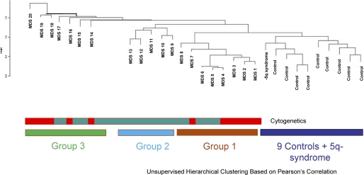
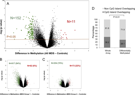
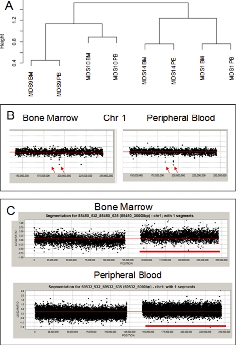
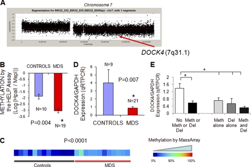
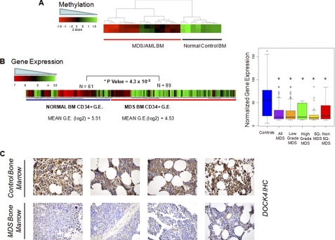
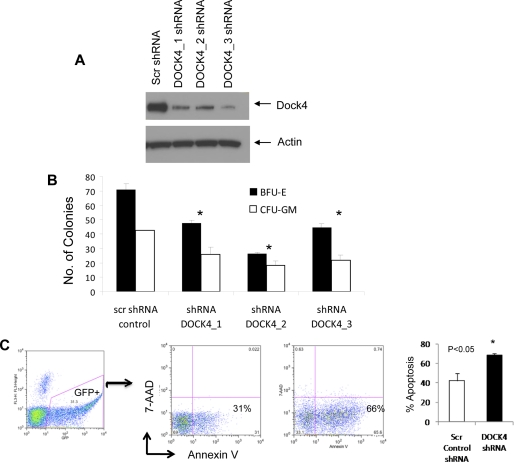
Myelodysplastic syndromes (MDS) are characterized by abnormal and dysplastic maturation of all blood lineages. Even though epigenetic alterations have been seen in MDS marrow progenitors, very little is known about the molecular alterations in dysplastic peripheral blood cells. We analyzed the methylome of MDS leukocytes by the HELP assay and determined that it was globally distinct from age-matched controls and was characterized by numerous novel, aberrant hypermethylated marks that were located mainly outside of CpG islands and preferentially affected GTPase regulators and other cancer-related pathways. Additionally, array comparative genomic hybridization revealed that novel as well as previously characterized deletions and amplifications could also be visualized in peripheral blood leukocytes, thus potentially reducing the need for bone marrow samples for future studies. Using integrative analysis, potentially pathogenic genes silenced by genetic deletions and aberrant hypermethylation in different patients were identified. DOCK4, a GTPase regulator located in the commonly deleted 7q31 region, was identified by this unbiased approach. Significant hypermethylation and reduced expression of DOCK4 in MDS bone marrow stem cells was observed in two large independent datasets, providing further validation of our findings. Finally, DOCK4 knockdown in primary marrow CD34(+) stem cells led to decreased erythroid colony formation and increased apoptosis, thus recapitulating the bone marrow failure seen in MDS. These findings reveal widespread novel epigenetic alterations in myelodysplastic leukocytes and implicate DOCK4 as a pathogenic gene located on the 7q chromosomal region.


Figures

FIGURE 1.
FIGURE 1.
Methylation profiling on peripheral blood leukocytes separates distinct subsets of MDS from normals. Methylation profiles generated by the HELP assay were used to cluster 21 MDS and 9 control samples by hierarchical clustering. The controls formed a cluster that was distinct from MDS samples. The MDS samples included two clusters (groups 1 and 2) of epigenetically similar samples with a greater amount of resemblance to controls. The remaining seven MDS samples demonstrated greater heterogeneity. No correlation with cytogenetics (normal represented as green and abnormal as red) was seen.
FIGURE 2.
FIGURE 2.
Majority of differentially methylated loci are hypermethylated in MDS leukocytes and reside outside of CpG islands. A volcano plot is shown demonstrating the difference in mean methylation between all MDS samples and controls on the x axis and the log of the p values between the means on the y axis. A two-tailed t test was used to calculate the p values. Significantly methylated loci with a log fold change in mean methylation are labeled in green, and significantly hypomethylated loci are labeled in red (A). Volcano plots for MDS subgroups 1 and 2 also reveal mostly hypermethylated loci with a variable number of hypomethylated loci. B and C, genomic position of every HpaII-amplifiable fragment on the HELP array was compared with the location of known CpG islands, and the fragments on the array were divided into two categories, those overlapping with these genomic elements and those not overlapping. To determine whether the differentially methylated genes between MDS and controls were enriched for either one of these types of elements, a proportions test was used to compare the relative proportion of the two types of HpaII fragments in the signature with the relative proportion on the array. Stacking bars are used to illustrate the finding of a significant enrichment for HpaII-amplifiable fragments not overlapping with CpG islands (D).
FIGURE 3.
FIGURE 3.
Array CGH can detect copy number variations in MDS leukocytes. Unsupervised clustering of copy number analysis reveals similarity between matched marrow (BM) and peripheral blood (PB) samples from the same patient (A). Array CGH plots of chromosome 1 reveal small deletions seen in both bone marrow and peripheral blood samples from one patient (B). In another patient with amplification of the short arm of chromosome 1 in bone marrow cells, the amplification is also seen in peripheral blood (C).
FIGURE 4.
FIGURE 4.
Integrative analysis reveals DOCK4 to be silenced by both deletion and hypermethylation in MDS. The aCGH plot of chromosome 7 from an MDS patient with 7q deletion shows the location of the DOCK4 gene (A). Mean methylation from the HELP assay (depicted by log2(HpaII/MspI)) is significantly higher in MDS samples as evident from a more negative value (two-tailed t test) (B). Methylation analysis of the DOCK4 promoter by MassArray analysis reveals greater methylation in MDS samples as depicted in the heat map (C). quantitative RT-PCR was performed on RNA from MDS leukocytes and control samples and showed a significantly reduced expression in MDS (D). Means ± S.E. with p value were calculated by two-tailed t test. Expression of DOCK4 was evaluated in MDS samples with or without deletions (Del) and promoter methylation (Meth) and shows significant reduction with either genetic or epigenetic silencing. Means ± S.E. with p value were calculated by two-tailed t test (E).
FIGURE 5.
FIGURE 5.
Validation in independent cohorts demonstrate reduction in DOCK4 in marrow samples from MDS/AML. Methylation values obtained from the HELP assay performed on marrow (BM) samples in an independent cohort of patients (38) show hypermethylation of the promoter in MDS/AML samples (A). Gene expression values from various studies on MDS and normal bone marrow-derived CD34+ cells were obtained and normalized. Mean expression of DOCK4 was significantly reduced in 89 MDS cases when compared with 61 controls (two-tailed t test) (B, left panel); box plots of MDS subtypes show significantly reduced levels of DOCK4 in all subtypes of MDS (B, right panel). Bone marrow biopsy samples were stained with DOCK4 antibody and show decreased expression in four representative cases of MDS when compared with controls (C).
FIGURE 6.
FIGURE 6.
DOCK4 knockdown leads to ineffective hematopoiesis in vitro. DOCK4 protein expression was reduced by three lentiviral mediated shRNA constructs (A). Primary bone marrow CD34+ stem cells with DOCK4 shRNAs produced fewer erythroid (erythroid burst-forming units (BFU-E)) and myeloid (CFU-GM) colonies (means ± S.E.; t test, p value< 0.05) (B). DOCK4 shRNA was able to increase apoptosis significantly in GFP+-sorted CD34+ cells (t test, p value< 0.05). Three independent experiments shown as means ± S.E. (C).

References

    1. Pellagatti A., Jädersten M., Forsblom A. M., Cattan H., Christensson B., Emanuelsson E. K., Merup M., Nilsson L., Samuelsson J., Sander B., Wainscoat J. S., Boultwood J., Hellström-Lindberg E. (2007) Proc. Natl. Acad. Sci. U.S.A. 104, 11406–11411 - PMC - PubMed
    1. Pellagatti A., Cazzola M., Giagounidis A. A., Malcovati L., Porta M. G., Killick S., Campbell L. J., Wang L., Langford C. F., Fidler C., Oscier D., Aul C., Wainscoat J. S., Boultwood J. (2006) Blood 108, 337–345 - PubMed
    1. Jones P. A., Baylin S. B. (2002) Nat. Rev. Genet. 3, 415–428 - PubMed
    1. Christiansen D. H., Andersen M. K., Pedersen-Bjergaard J. (2003) Leukemia 17, 1813–1819 - PubMed
    1. Aggerholm A., Holm M. S., Guldberg P., Olesen L. H., Hokland P. (2006) Eur. J. Haematol. 76, 23–32 - PubMed

Publication types

MeSH terms