Review
. 2011 May-Jun;5(3):193-7.
doi: 10.4161/chan.5.3.15765. Epub 2011 May 1.

Pannexin channels are not gap junction hemichannels

Affiliations
Review

Pannexin channels are not gap junction hemichannels

Gina E Sosinsky et al. Channels (Austin). 2011 May-Jun.

Abstract

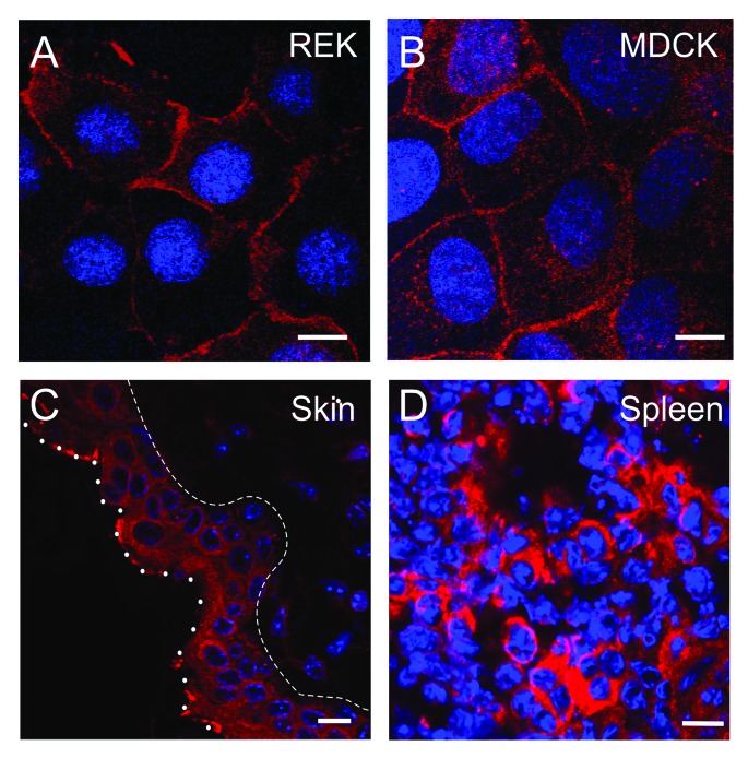
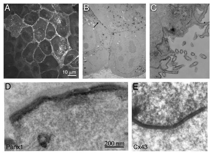
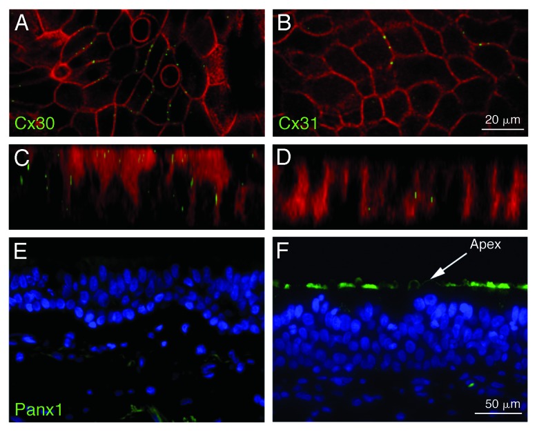
Pannexins, a class of membrane channels, bear significant sequence homology with the invertebrate gap junction proteins, innexins and more distant similarities in their membrane topologies and pharmacological sensitivities with the gap junction proteins, connexins. However, the functional role for the pannexin oligomers, or pannexons, is different from connexin oligomers, the connexons. Many pannexin publications have used the term "hemichannels" to describe pannexin oligomers while others use the term "channels" instead. This has led to confusion within the literature about the function of pannexins that promotes the idea that pannexons serve as gap junction hemichannels and thus have an assembly and functional state as gap junctional intercellular channels. Here we present the case that unlike the connexin gap junction intercellular channels, so far, pannexin oligomers have repeatedly been shown to be channels that are functional in single membranes, but not as intercellular channel in appositional membranes. Hence, they should be referred to as channels and not hemichannels. Thus, we advocate that in the absence of firm evidence that pannexins form gap junctions, the use of the term "hemichannel" be discontinued within the pannexin literature.


Figures

None
Figure 1. Pannexins are predicted to be tetra-spanning membrane proteins with both the amino and carboxy termini exposed to the cytoplasm. Panx1 and Panx3 have N-linked glycosylation sites at N254 and N71, respectively (red circles), while Panx2 has a predicted N-linked glycosylation site at N86 (orange circle). Black circles denote the four conserved cysteines.
None
Figure 2. Subcellular localization of Panx1 varies in cells and tissues. Rat epidermal keratinocytes (REK) (A) overexpressing Panx1 show a similar cell surface profile to endogenous Panx1 in Madin-Darby canine kidney cells (MDCK) (B) as revealed via immunolabeling for Panx1 (red). Panx1 exhibits a diffused cellular phenotype in mouse epidermis (between the dashed and dotted lines) (C) not unlike the distribution of Panx1 in thin sections of mouse spleen (D). Nuclei are stained with Hoechst (blue). Bars = 10 μm.
None
Figure 3. Correlative light and electron microscopy of tagged Panx1 in MDCK cells. Panx1 was tagged with a tetracysteine domain, labeled with ReAsH and photo-oxidized after fluorescence imaging. ReAsH fluorescence (A) Low magnification EM of the same cells (B and C). Higher power EM of area denoted by an arrow in (B), showed staining at appositional areas between cells, where a clear separation of the two plasma membranes are observed (D). Photo-oxidized Cx43-tetracysteine/ReAsH gap junction is shown for comparison (E). (D and E) are displayed with the same magnification. (Adapted from Boassa et al. (2007). © The American Society for Biochemistry and Molecular Biology).
None
Figure 4. Localization of connexins and Panx1 in airway epithelial cells. These cells express the two connexins, Cx30 and Cx31, which yield the typical punctate staining of gap junctions at the basolateral membranes of contacting cells (A and B). Z-stacks (C and D) do not reveal staining of connexins at the apical membrane. In contrast, staining for Panx1 is restricted to the apical membrane where ATP is released from these cells (E and F). No staining at the basolateral membrane can be detected. (A–D) are from Wiszniewski et al. (2007), with permission from Elsevier. (E and F) are adapted from Ransford et al. (2009), reprinted with permission of the American Thoracic Society. © Copyright American Thoracic Society.

Similar articles

Cited by

References

    1. Baranova A, Ivanov D, Petrash N, Pestova A, Skoblov M, Kelmanson I, et al. The mammalian pannexin family is homologous to the invertebrate innexin gap junction proteins. Genomics. 2004;83:706–16. doi: 10.1016/j.ygeno.2003.09.025. - DOI - PubMed
    1. Bruzzone R, Hormuzdi SG, Barbe MT, Herb A, Monyer H. Pannexins, a family of gap junction proteins expressed in brain. Proc Natl Acad Sci U S A. 2003;100:13644–9. doi: 10.1073/pnas.2233464100. - DOI - PMC - PubMed
    1. Panchin Y, Kelmanson I, Matz M, Lukyanov K, Usman N, Lukyanov S. A ubiquitous family of putative gap junction molecules. Curr Biol. 2000;10:R473–4. doi: 10.1016/S0960-9822(00)00576-5. - DOI - PubMed
    1. Penuela S, Bhalla R, Gong XQ, Cowan KN, Celetti SJ, Cowan BJ, et al. Pannexin 1 and pannexin 3 are glycoproteins that exhibit many distinct characteristics from the connexin family of gap junction proteins. J Cell Sci. 2007;120:3772–83. doi: 10.1242/jcs.009514. - DOI - PubMed
    1. Maeda S, Nakagawa S, Suga M, Yamashita E, Oshima A, Fujiyoshi Y, et al. Structure of the connexin 26 gap junction channel at 3.5 A resolution. Nature. 2009;458:597–602. doi: 10.1038/nature07869. - DOI - PubMed

LinkOut - more resources