Review
. 2011 May;127(5):105e-118e.
doi: 10.1097/PRS.0b013e31820cf556.

Adult peripheral nerve disorders: nerve entrapment, repair, transfer, and brachial plexus disorders

Affiliations
Review

Adult peripheral nerve disorders: nerve entrapment, repair, transfer, and brachial plexus disorders

Ida K Fox et al. Plast Reconstr Surg. 2011 May.

Abstract

Learning objectives: After reading this article, the participant should be able to: 1. Describe the pathophysiologic bases for nerve injury and how they apply to patient evaluation and management. 2. Recognize the wide variety of injury patterns and associated patient complaints and physical findings associated with peripheral nerve pathology. 3. Evaluate and recommend further tests to aid in defining the diagnosis. 4. Specify treatment options and potential risks and benefits.

Summary: Peripheral nerve disorders comprise a gamut of problems, ranging from entrapment neuropathy to direct open traumatic injury and closed brachial plexus injury. The pathophysiology of injury defines the patient's symptoms, examination findings, and treatment options and is critical to accurate diagnosis and treatment. The goals of treatment include management of the often associated pain and improvement of sensory and motor function. Understanding peripheral nerve anatomy is critical to adopting novel nerve transfer procedures, which may provide superior options for a variety of injury patterns.


Figures
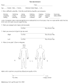
Figure 1
Figure 1
Pain Questionnaire. We find this diagram quite useful for localizing and describing the symptoms related to nerve injury particularly in nerve entrapment syndromes.
Figure 2
Figure 2. Spinal Accessory to Suprascapular Motor Nerve Transfer
In this transfer, spinal accessory (Cranial Nerve XI) motor fibers are re-routed and repaired to the non-functional suprascapular nerve. This is particularly useful in patients with brachial plexus injuries with dysfunction due to nerve root level avulsions rendering the musculature innervated by the suprascapular nerve non-functional. A posterior transverse incision is made just cephalad to the spine of the scapula and the nerves are isolated on the underside of the trapezius (spinal accessory donor nerve) and at the suprascapular notch (suprascapular recipient nerve). The donor nerve is cut as far distally as possible and the recipient is cut proximally after release at the notch to allow for easy, tension-free repair. (Reproduced with permission, Mackinnon SE and Colbert, SH, Nerve Transfers in the Hand and Upper Extremity Surgery, Techniques in Hand and Upper Extremity Surgery, 12: 20–33, 2008.)
Figure 3
Figure 3. Radial to Axillary Motor Nerve Transfer
In this transfer, radial nerve branch to triceps medial head muscle motor fibers are rerouted and repaired to the non-functional axillary nerve branch to the deltoid muscle. This is particularly useful in patients with brachial plexus injuries with dysfunction due to nerve root level avulsions rendering the musculature innervated by the deltoid nerve nonfunctional. A curvilinear incision is made over the posterior shoulder and the nerves are isolated at the posterior arm between the triceps heads (medial triceps nerve branch of radial nerve donor nerve) and in the quadrangular space (axillary recipient nerve). The donor nerve is cut as far distally as possible and the recipient is cut proximally to allow for easy, tension-free repair. The donor nerve fibers can be repaired to preferentially reinnervate the recipient motor fibers by neurolysing out and excluding the sensory component of the axillary nerve. (Reproduced with permission, Mackinnon SE and Colbert, SH, Nerve Transfers in the Hand and Upper Extremity Surgery, Techniques in Hand and Upper Extremity Surgery, 12: 20–33, 2008.)
Figure 4
Figure 4. Median to Ulnar Motor and Sensory Nerve Transfers
In this transfer, anterior interosseous (median) nerve branch to pronator quadratus muscle motor fibers are re-routed and repaired to the non-functional ulnar nerve deep motor branch. This is particularly useful in adult patients with very proximal ulnar nerve injuries. Although direct exploration and repair may allow restoration of ulnar innervated extrinsic musculature, intrinsic function will not be restored due to the length of time required. By the time regenerating fibers reach the hand level intrinsic muscle fibers, that muscle will be non-functional. By use of more distal transfers, anticlaw procedures can be avoided. In addition, critical areas of sensation can be restored in a more timely fashion. In figures a and b, the motor branches are isolated, transected and repaired to effect the motor transfer. In figures c and d, the sensory branches are likewise dissected out and repaired to restore sensation to the ulnar border of the hand--in this case sensation to the non-critical third web space is sacrificed. (Reproduced with permission, Mackinnon SE and Colbert, SH, Nerve Transfers in the Hand and Upper Extremity Surgery, Techniques in Hand and Upper Extremity Surgery, 12: 20–33, 2008.)

Similar articles

Cited by

References

    1. Mackinnon SE, Dellon AL. Surgery of the Peripheral Nerve. New York: Thieme Medical Publishers, Inc; 1988.
    1. Sunderland S. A classification of peripheral nerve injuries producing loss of function. Brain. 1951;74:491–516. - PubMed
    1. Mackinnon SE, Novak CB. Compression Neuropathies. In: Green DP, Hotchkiss RN, Pederson WC, et al., editors. Green’s Operative Hand Surgery. 5. Vol. 1. Philadelphia: Elsevier, Inc; 2005. pp. 999–1045.
    1. Strauch B, Lang A. The ten test revisited. Plast Reconstr Surg. 2003;112:593–594. - PubMed
    1. Strauch B, Lang A, Ferder M, et al. The ten test. Plast Reconstr Surg. 1997;99:1074–1078. - PubMed

Publication types

MeSH terms