

Assessment of fetal well-being in cattle by ultrasonography in normal, high-risk, and cloned pregnancies
- PMID: 21532817
- PMCID: PMC3022448
Assessment of fetal well-being in cattle by ultrasonography in normal, high-risk, and cloned pregnancies
Abstract
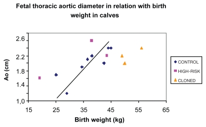
This study determined ultrasonographic parameters of fetuses and uterine adnexa in late pregnancy in normal, cloned, and high-risk pregnancies in relation to perinatal and neonatal outcome. Ten cows with normal pregnancies (CONTROL, mean pregnancy length 273 d), 10 sick cows with potentially compromised pregnancies (HIGH-RISK, mean pregnancy length 267 d), and 10 heifers with cloned pregnancies (CLONED, mean pregnancy length 274 d) were examined at more than 260 d of gestation. There was no difference in mean fetal heart rates among the groups. The cloned calves were heavier (57 ± 8 kg) than calves from CONTROL group (36 ± 7 kg), and calves from HIGH-RISK group (37 ± 13 kg) (P = 0.003). The diameter of the thoracic aorta was positively correlated (R = 0.62) with fetal birth weight in the CONTROL group (P = 0.01). Fetal activity was not associated with survival. The results suggest that transabdominal ultrasonographic assessment of the fetal well-being may serve as a potential tool for evaluation of the fetoplacental unit.
Évaluation par échographie du bien-être fœtal chez le bétail pour des gestations normales, à risque élevé et celles d’animaux clonés. Cette étude a déterminé les paramètres échographiques des fœtus et des annexes utérines à la fin de la gestation pour des gestations normales et à risque élevé par rapport aux résultats périnataux et néonataux. Dix vaches avec des gestations normales (TÉMOIN, durée moyenne de la gestation de 273 jours), 10 vaches malades avec des gestations potentiellement compromises (RISQUE ÉLEVÉ, durée moyenne de gestation de 267 jours) et 10 génisses avec des grossesses d’animaux clonés (CLONÉS, durée moyenne de gestation de 274 jours) ont été examinées à plus de 260 jours de gestation. Il n’y avait aucune différence au niveau des rythmes cardiaques fœtaux moyens parmi les groupes. Les veaux clonés étaient plus lourds (57 ± 8 kg) que les veaux du groupe TÉMOIN (36 ± 7 kg) et que les veaux du groupe à RISQUE ÉLEVÉ (37 ± 13 kg) (P = 0,003). Le diamètre de l’aorte thoracique présentait une corrélation positive (R = 0,62) avec le poids fœtal à la naissance du groupe TÉMOIN (P = 0,01). L’activité fœtale n’était pas associée à la survie. Les résultats suggèrent qu’une évaluation transabdominale du bien-être fœtal peut servir d’outil potentiel pour l’évaluation de l’unité fœto-placentaire.
(Traduit par Isabelle Vallières)
Figures
References
-
- Lerner JP. Fetal growth and well-being. Obstet Gynecol Clin North Am. 2004;31:159–176. - PubMed
-
- Manning FA. The fetal biophysical profile score. Obstet Gynecol Clin North Am. 1999;26:557–577. - PubMed
-
- Manning FA. Ultrasonography in perinatal medicine. In: Avery GB, editor. Neonatalogy: Pathophysiology and Management of the Newborn. 3rd ed. Philadelphia: Lippincott; 1987. pp. 110–129.
-
- Reef VB, Vaala WE, Worth LT, et al. Ultrasonographic evaluation of the fetus and intrauterine environment in healthy mares during late gestation. Vet Radiol Ultrasound. 1995;36:533–541.
Publication types
MeSH terms
LinkOut - more resources
Full Text Sources