

A rare case of multiple segmental eccrine spiradenomas
- PMID: 21532877
- PMCID: PMC3084607
A rare case of multiple segmental eccrine spiradenomas
Abstract
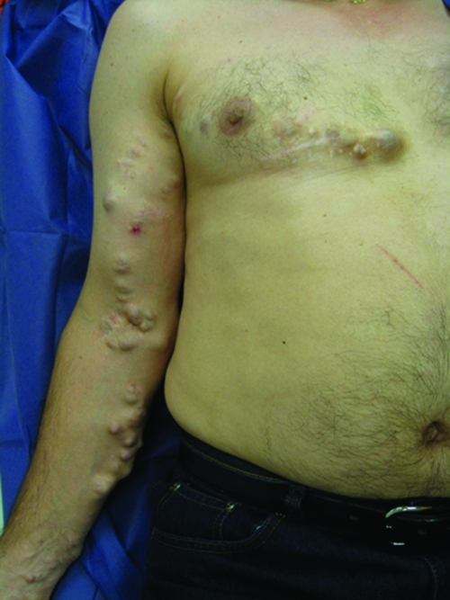
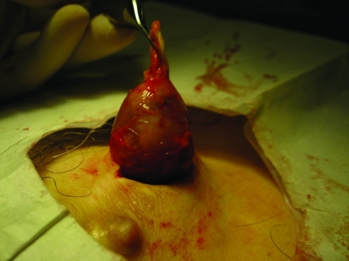
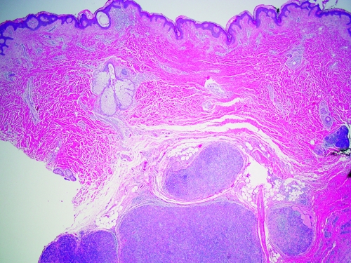
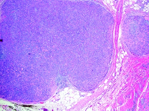
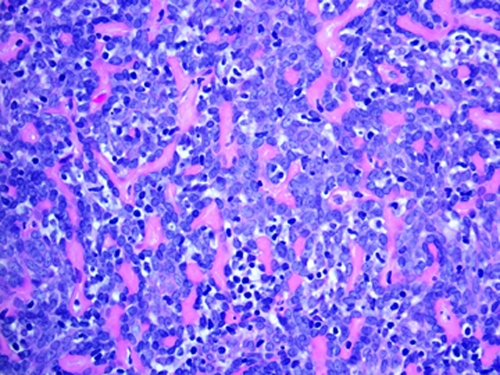
Eccrine spiradenoma is a benign adnexal neoplasm that has been historically designated as a tumor of eccrine differentiation, although current reconsideration indicates an apocrine process. It usually presents on the trunk and extremities as a tender dermal or subcutaneous papule or nodule frequently with a pink or blue hue. The clinical picture is often not distinct and biopsy is required for diagnosis. Eccrine spiradenoma can present in a variety of ways, including as tumors arranged in zosteriform/dermatomal and/or blaschkoid distributions, often precluding a straightforward diagnosis. Proper diagnosis of eccrine spiradenoma is important due to the occurrence of potentially life-threatening malignant transformation. This article illustrates a rare presentation of eccrine spiradenoma with a concise review for the dermatologist.
Figures
References
-
- Kersting DW, Helwig EB. Eccrine spiradenoma. AMA Arch Derm. 1956;73(3):199–227. - PubMed
-
- Nath AK, Kumari R, Thappa DM. Eccrine spiradenoma with chondroid syringoma in Blaschkoid distribution. Indian J Dermatol Venereol Leprol. 2009;75(6):600–602. - PubMed
-
- Bedlow AJ, Cook MG, Kurwa A. Extensive naevoid eccrine spiradenoma. Br J Dermatol. 1999;140(1):154–157. - PubMed
-
- Ekmekci TR, Koslu A, Sakiz D. Congenital blaschkoid eccrine spiradenoma on the face. Eur J Dermatol. 2005;15(2):73–74. - PubMed
-
- Gupta S, Jain VK, Singh U, Gupta S. Multiple eccrine spiradenoma in zosteriform distribution in a child. Ped Dermatol. 2000;17(5):384–386. - PubMed
Publication types
LinkOut - more resources
Full Text Sources