. 2011 May;13(5):461-71.
doi: 10.1593/neo.101744.

Identification of an exon 4-deletion variant of epidermal growth factor receptor with increased metastasis-promoting capacity

Affiliations

Identification of an exon 4-deletion variant of epidermal growth factor receptor with increased metastasis-promoting capacity

Hai Wang et al. Neoplasia. 2011 May.

Abstract

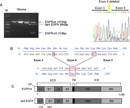
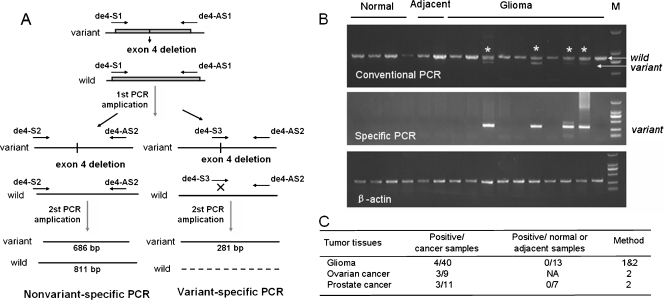
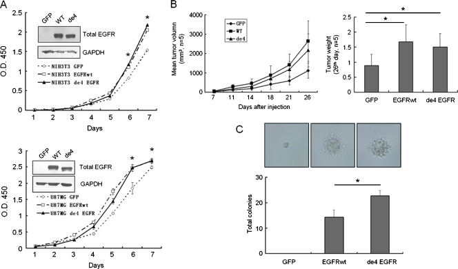
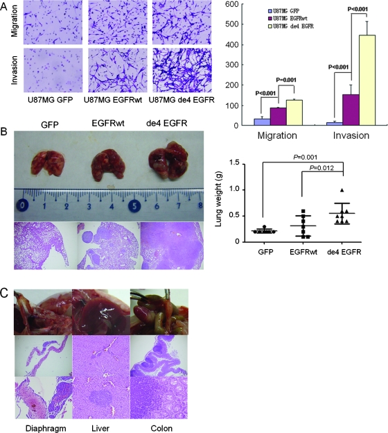
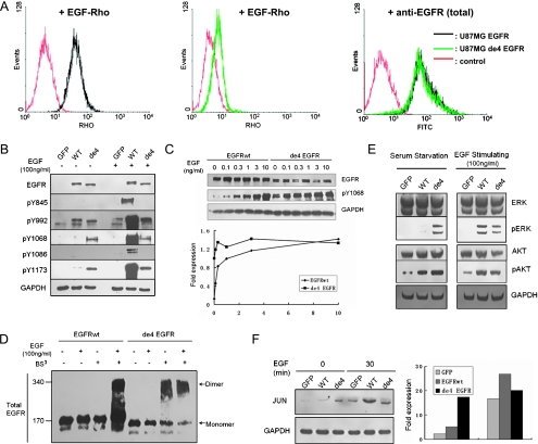
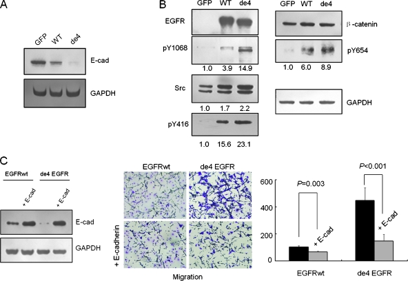
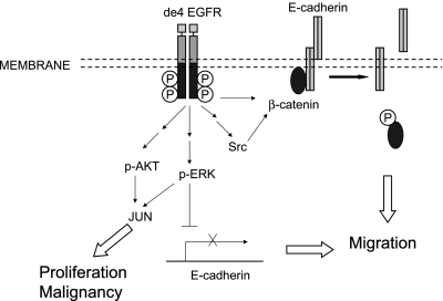
Several types of epidermal growth factor receptor (EGFR) gene alternations have been observed in human tumors. Here we present a novel EGFR variant with aberrant splicing of exon 4 (named as de4 EGFR). Variant-specific polymerase chain reaction showed that de4 EGFR was expressed in some glioma (4/40), prostate cancer (3/11), and ovarian cancer (3/9) tissues but not in tissues adjacent to tumors or normal tissues. de4 EGFR displayed an enhanced transformation and a higher metastasis-promoting capacity in comparison to wild-type EGFR. With minimal EGF-binding activity, de4 EGFR underwent ligand-independent autophosphorylation and self-dimerization. Moreover, in serum-starved condition, de4 EGFR expression in U87 MG cells significantly upregulated the extracellular signal-regulated kinase and AKT phosphorylation and expression of JUN and Src. Importantly, E-cadherin expression was barely detectable in the U87 MG cells expressing de4 EGFR and restored expression of E-cadherin in these cells inhibited their metastatic behaviors. Taken together, we identified a novel EGFR variant with increased metastasis-promoting activity that may become a promising new target for cancer therapy.


Figures

Figure 1
Figure 1
Identification of de4 EGFR. (A) The discovery of de4 EGFR in glioma. The amplicon and the corresponding length of each EGFR variant are shown by an arrow. The right panel shows the determined sequencing result of de4 EGFR. (B) Salient features of de4 EGFR. The in-frame splicing removes exon 4 and creates a novel glycine residue at the splice junction. (C) Schematic representation of de4 EGFR variant. Deleted amino acid numbers are indicated. ECD indicates extracellular domain (S1, L1, S2, and L2 are subdomains); ICD, intracellular domain; RD, regulatory domain; SP, signal peptide; TKD, tyrosine kinase domain; TM, transmembrane.
Figure 2
Figure 2
de4 EGFR is observed in multiple cancer tissues. (A) Schematic of the PCR method used to detect the deletion of exon 4 in the EGFR gene. (B) Detection of EGFRwt and de4 EGFR in gliomas by conventional and variant-specific PCR. (Partial results) The variant amplicons shown here were sequenced. β-Actin served as an internal control. (C) Summary of the occurrence of de4 EGFR in several cancer tissues. Method 1: detection by conventional PCR. Method 2: detection by variant-specific PCR. NA indicates not applicable.
Figure 3
Figure 3
de4 EGFR promotes proliferation and transformation. (A) Growth curve showing the mitogenic activities of U87MG (bottom) and NIH/3T3 (top) transfectants in vitro. Inset: immunoblot analysis of whole lysates of the NIH/3T3 and U87MG transfectants using an anti-EGFR antibody. (B) Both EGFRwt and de4 EGFR promoted the proliferation of U87MG in vivo (n = 5). The right panel shows a comparison of tumor weight. (C) Colony formation in soft agar. NIH/3T3 cell derivatives (300/well) were cultured for 3 weeks. de4 EGFR transfected cells form the most colonies. *P < .05, compared with the control. Error bars, SD. Photomicrographs of colonies are representative. Magnification, x400. The normality of each data set was confirmed using the Levene test. Statistical data were evaluated using an ANOVA. Comparisons between two means were evaluated using the LSD method.
Figure 4
Figure 4
de4 EGFR promotes invasion and metastasis in vitro and in vivo. (A) de4 EGFR enhances migration and invasion of U87MG in vitro (quantified on the right). Magnification, x100. Representative experiments are shown in triplicate along with SD. (B) de4 EGFR promotes more severe spontaneous metastases after subcutaneous inoculation of U87MG transfectants. Left: Comparison of lung lesions caused by three transfectants and hematoxylin and eosin staining of formalin-fixed lung sections. Magnification, x40. Right: Comparison of lung weight (tumor burden). Error bars, SD (n = 7/7/8). The normality of each data set was confirmed using the Levene test. Statistical data were evaluated using an ANOVA. Comparisons between two means were evaluated using the LSD method. (C) U87MG-de4 EGFR induced extrapulmonary metastatic foci in the diaphragm (left), liver (middle), and colon (right).
Figure 5
Figure 5
Oncogenic signaling of de4 EGFR. (A) Flow cytometry analysis for EGF-Rho binding affinity. Reactivity was tested on U87MG-EGFR (left) and U87-de4 EGFR (middle). Expression of total EGFR was assessed first (right). Autofluorescence blanks are also shown (red). (B) Tyrosine phosphorylation of EGFRwt and de4 EGFR treated with or without EGF for 8 minutes. Y1068 and Y1173 were phosphorylated in the presence of de4 EGFR in the absence of EGF activation. GAPDH served as an internal control. (C) U87MG transfectants expressing either EGFRwt or de4 EGFR were incubated with increasing EGF concentrations. The phosphorylation of Y1068 was analyzed by immunoblot analysis with the indicated antibodies. Phospho-EGFR levels were quantified according to band intensities. (D) Dimerization analysis of EGFRwt and de4 EGFR. Cells were treated with or without EGF (100 ng/ml; 8 minutes) and incubated with or without a covalent cross-linking reagent, BS3 (2mM, 20 minutes). EGFR dimers and monomers are indicated, respectively. (E) General signaling pathways relating to EGFR. ERK and AKT are activated in the presence of de4 EGFR. GAPDH served as an internal control. (F) The relative levels of JUN were determined by immunoblot analysis. The bands were quantified according to band intensities. GAPDH served as an internal control.
Figure 6
Figure 6
Reduction of E-cadherin-mediated cell-cell adhesion contributes to the increased migratory capacity of cell lines expressing de4 EGFR. (A) E-cadherin expression levels in three U87MG transfectants. (B) Activation of EGFR and overexpressed Src induced phosphorylation of β-catenin. Numbers indicate normalized intensities of bands. Cells were serum-starved for 8 hours and then harvested. (C) Exogenous E-cadherin was successfully transfected into U87MG-EGFR and U87MG-de4 EGFR. Enhanced E-cadherin expression resulted in decreased migratory capacity of U87MG-EGFR and U87MG-de4 EGFR. The normality of each data set was confirmed using the Levene test. Statistical data were conducted using the t test method.
Figure 7
Figure 7
Model showing how de4 EGFR promotes cell malignancy. Unlike EGFRwt, de4 EGFR shows basal phosphorylation independent of EGF stimulation. de4 EGFR likely enhances proliferation and transformation through constitutive activation of ERK/AKT and upregulation of the expression level of JUN in the absence of ligand stimulation. Conversely, de4 EGFR drives migration by down-regulation of E-cadherin-mediated cell-cell adhesion through two different mechanisms. One involves constitutive activation of ERK and thus inhibits E-cadherin expression. The other pathway includes β-catenin phosphorylation induced by overexpressed Src and basal activation of EGFR. This leads to the destruction of E-cadherin/catenin adhesive complexes and hence facilitating cell migration.

Similar articles

Cited by

References

    1. Xue C, Wyckoff J, Liang F, Sidani M, Violini S, Tsai K, Zhang Z, Sahai E, Condeelis J, Segall JE. Epidermal growth factor receptor overexpression results in increased tumor cell motility in vivo coordinately with enhanced intravasation and metastasis. Cancer Res. 2006;66:192–197. - PubMed
    1. Pandiella A, Lehvaslaiho H, Magni M, Alitalo K, Meldolesi J. Activation of an EGFR/neu chimeric receptor: early intracellular signals and cell proliferation responses. Oncogene. 1989;4:1299–1305. - PubMed
    1. Liang K, Ang KK, Milas L, Hunter N, Fan Z. The epidermal growth factor receptor mediates radioresistance. Int J Radiat Oncol Biol Phys. 2003;57:246–254. - PubMed
    1. Ho R, Minturn JE, Hishiki T, Zhao H, Wang Q, Cnaan A, Maris J, Evans AE, Brodeur GM. Proliferation of human neuroblastomas mediated by the epidermal growth factor receptor. Cancer Res. 2005;65:9868–9875. - PubMed
    1. Modjtahedi H, Essapen S. Epidermal growth factor receptor inhibitors in cancer treatment: advances, challenges and opportunities. Anticancer Drugs. 2009;20:851–855. - PubMed

Publication types

MeSH terms

Substances