. 2011 May;13(5):472-82.
doi: 10.1593/neo.101748.

p38γ mitogen-activated protein kinase contributes to oncogenic properties maintenance and resistance to poly (ADP-ribose)-polymerase-1 inhibition in breast cancer

Affiliations

p38γ mitogen-activated protein kinase contributes to oncogenic properties maintenance and resistance to poly (ADP-ribose)-polymerase-1 inhibition in breast cancer

Fanyan Meng et al. Neoplasia. 2011 May.

Abstract

p38γ MAPK, one of the four members of p38 mitogen-activated protein kinases (MAPKs), has previously been shown to harbor oncogenic functions. However, the biologic function of p38γ MAPK in breast cancer has not been well defined. In this study, we have shown that p38γ MAPK is overexpressed in highly metastatic human and mouse breast cancer cell lines and p38γ MAPK expression is preferentially associated with basal-like and metastatic phenotypes of breast tumor samples. Ectopic expression of p38γ MAPK did not lead to an increase in oncogenic properties in vitro in most tested mammary epithelial cells. However, knockdown of p38γ MAPK expression resulted in a dramatic decrease in cell proliferation, colony formation, cell migration, invasion in vitro and significant retardation of tumorigenesis, and long-distance metastasis to the lungs in vivo. Moreover, knockdown of p38γ MAPK triggered the activation of AKT signaling. Inhibition of this feedback loop with various PI3K/AKT signaling inhibitors facilitated the effect of targeting p38γ MAPK. We further found that overexpression of p38γ MAPK did not promote cell resistance to chemotherapeutic agents doxorubicin and paclitaxel but significantly increased cell resistance to PJ-34, a DNA damage agent poly (ADP-ribose)-polymerase-1 (PARP) inhibitor in vitro and in vivo. Finally, we identified that p38γ MAPK overexpression led to marked cell cycle arrest in G(2)/M phase. Our study for the first time clearly demonstrates that p38γ MAPK is a promising target for the design of targeted therapies for basal-like breast cancer with metastatic characteristics and for overcoming potential resistance against the PARP inhibitor.


Figures

Figure 1
Figure 1
Expression of p38γ MAPK in breast cancer cell lines and breast tumor tissues. (A and B) Expression of p38γ MAPK in human (A) and mouse (B) highly metastatic cell lines. Top panel shows RT-PCR results. Lower panel shows results of Western blot analysis. (C) Expression of p38γ MAPK is preferentially associated with basal-like breast cancer (P = .006). Group A is basal-like breast tumor (116 cases); group B is non-basal-like breast tumor (83 cases). (D) IHC assay shows that p38γ MAPK is significantly overexpressed in the tumor samples with lymph node and distant metastasis (P = .001). Group A (n = 25) includes tumor samples with lymph node and distant metastasis. Group B (n = 50) includes tumor samples without metastasis and normal tissue.
Figure 2
Figure 2
Effect of knockdown of p38γ MAPK on oncogenic properties in vitro. (A) Knockdown of p38γ MAPK expression in 4T1 cells significantly inhibits cell proliferation. Top panel shows five shRNA clones that differentially inhibit the expression of p38γ MAPK in 4T1 cells. β-Actin is a protein-loading control. Lower panel shows two clones with knockdown of p38γ MAPK expression that have inhibited cell proliferation compared with parental 4T1 cell and 4T1 with nontarget control in 7 days. (B, C, and D) Knockdown of p38γ MAPK expression in 4T1 cells significantly inhibits colony formation (B), cell migration (C), and invasion (D). Two clones (sh1 and sh2) with knockdown of p38γ MAPK expression and one clone (sh3) without significant p38γ MAPK expression change were used to compare with parental 4T1 cell and 4T1 with nontarget control.
Figure 3
Figure 3
Effect of knockdown of p38γ MAPK expression in tumorigenesis and distal metastasis in vivo. (A and B) Two clones with knockdown p38γ MAPK expression (p38γ-sh1 and p38γ-sh2) show inhibited tumor burden in vivo (A) and reduced lung metastasis (B) compared with parental 4T1 cell, 4T1 with nontarget control, and 4T1 cells without significant p38γ MAPK expression change (p38γ-sh3). Ten mice were used in each group. (C) Representative hematoxylin and eosin staining pictures of lung sections from mice injected with 4T1-derived cells (magnification, x100 and x200). The origin of the lung section is shown on the right of the panels.
Figure 4
Figure 4
Effect of ectopic expression of p38γ MAPK on oncogenic properties of MCF7 cells in vitro and in vivo. (A) Ectopic expression of p38γ MAPK in MCF7 cells has no effect on cell proliferation. The top panel shows ectopic expression of p38γ MAPK in MCF7 cells. V5 antibody is used to detect p38γ MAPK. β-actin is a protein loading control. The lower panel shows cell proliferation of MCF7 cells expressing vector control and p38γ MAPK gene in 11 days. (B) Ectopic expression of p38γ MAPK in MCF7 cells does not lead to change in colony formation compared with MCF7 cells with vector control. (C) Ectopic expression of p38γ MAPK in MCF7 cells leads to an increase in cell migration (top panel) and cell invasion (low panel) compared with MCF7 cells with vector control. (D) Effect of ectopic expression of p38γ MAPK on tumorigenesis. The cells with ectopic p38γ MAPK expression show no significant difference in tumor burden in vivo compared with parental MCF7 cell with vector control. Eight mice were used in each group.
Figure 5
Figure 5
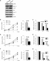
Effect of p38γ MAPK on cell signaling pathways. (A) Effect of p38γ MAPK on AKT and ERK signaling by Western blot analysis assay. Western blots show the changes in cell signaling when p38γ MAPK is knocked down in 4T1 cells. (B, D, and F) The effect of combination of knockdown of p38γ MAPK and various AKT signaling inhibitors on cell proliferation with different time courses. The right panels show the final comparison of the cell survival rate between nontarget clone and knockdown clone after treatment of different inhibitors. (C, E, and G) Effect of the combination of knockdown of p38γ MAPK and various AKT signaling inhibitors on colony formation with different doses. 4T1 cells with p38γ MAPK knockdown (MK) and nontarget clone (NT) were treated with AKT inhibitor (AKti-1/2) (B and C), PI3K inhibitor (LY294002) (D and E), and Src inhibitor (Dasatinib) (F and G).
Figure 6
Figure 6
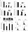
Effects of Dox and Pac on 67NR and MCF7 cells with or without p38γ MAPK overexpression. (A and D) Overexpression of p38γ MAPK in 67NR (A) and MCF7 (D) cells has no effect on the cell proliferation under the treatments with Dox and Pac. (B and E) Overexpression of p38γ MAPK in 67NR (B) and MCF7 (E) cells has no effect on the colony formation capability under the treatments with Dox. (C and F) Overexpression of p38γ MAPK in 67NR (C) and MCF7 (F) cells has no effect to the colony formation capability under the treatments with Pac.
Figure 7
Figure 7
Effect of ectopic expression of p38γ MAPK on cell resistance to the PARP inhibitor. (A and B) The 67NR cells (A) and HMLE cells (B) with ectopic expression of p38γ MAPK show significant resistance to PARP inhibitor PJ-34. The left panel shows the cell proliferation after PJ-34 treatment. The right panel shows the quantification of the clonogenic assay results. Different doses of PJ-34 are shown. (C) Effect of p38γ MAPK and PJ-34 treatment on tumorigenesis in vivo. Relative final tumor weights were shown after PJ-34 was used for 16 days. (D) Ectopic expression p38γ MAPK and PJ-34 treatment led to G2/M arrest in 67NR cells. 67NR cells with LacZ and p38γ MAPK expression were treated with DMSO (Untreated) and 15 µM PJ-34 for 2 days. Cell cycle was measured by flow cytometry assay.

Similar articles

Cited by

References

    1. Cuenda A, Rousseau S. p38 MAP-kinases pathway regulation, function and role in human diseases. Biochim Biophys Acta. 2007;1773:1358–1375. - PubMed
    1. Dhanasekaran DN, Johnson GL. MAPKs: function, regulation, role in cancer and therapeutic targeting. Oncogene. 2007;26:3097–3099. - PubMed
    1. Goedert M, Cuenda A, Craxton M, Jakes R, Cohen P. Activation of the novel stress-activated protein kinase SAPK4 by cytokines and cellular stresses is mediated by SKK3 (MKK6); comparison of its substrate specificity with that of other SAP kinases. EMBO J. 1997;16:3563–3571. - PMC - PubMed
    1. Han J, Lee JD, Bibbs L, Ulevitch RJ. A MAP kinase targeted by endotoxin and hyperosmolarity in mammalian cells. Science. 1994;265:808–811. - PubMed
    1. Jiang Y, Chen C, Li Z, Guo W, Gegner JA, Lin S, Han J. Characterization of the structure and function of a new mitogen-activated protein kinase (p38β) J Biol Chem. 1996;271:17920–17926. - PubMed

Publication types

MeSH terms

Substances

LinkOut - more resources