. 2011 Jan 1;58(1):27-37.
doi: 10.1016/j.dsr.2010.11.001.

The Inner Ear and its Coupling to the Swim Bladder in the Deep-Sea Fish Antimora rostrata (Teleostei: Moridae)

Affiliations

The Inner Ear and its Coupling to the Swim Bladder in the Deep-Sea Fish Antimora rostrata (Teleostei: Moridae)

Xiaohong Deng et al. Deep Sea Res 1 Oceanogr Res Pap. .

Abstract

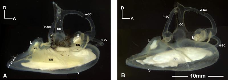
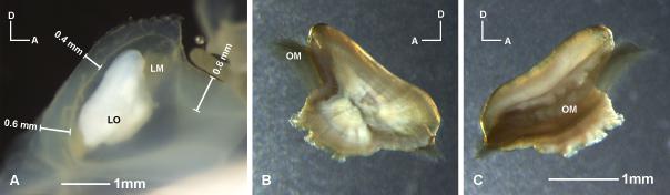
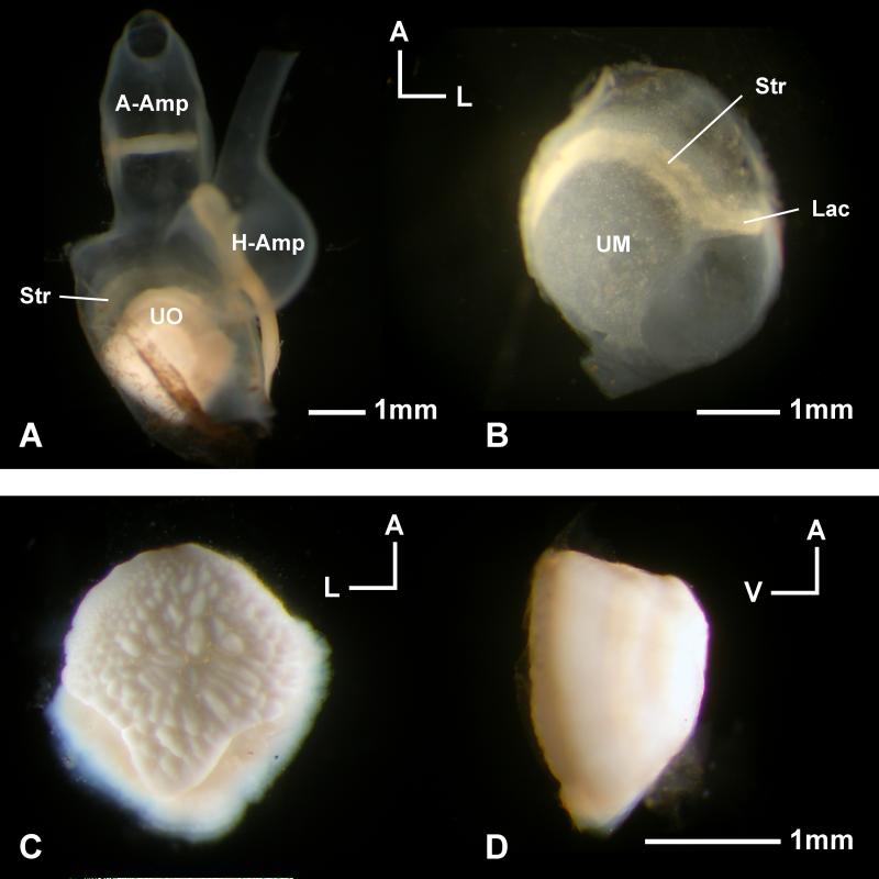
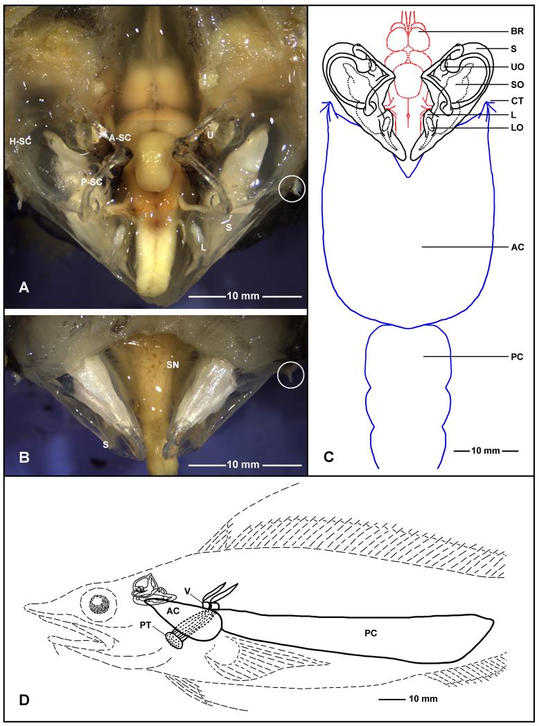
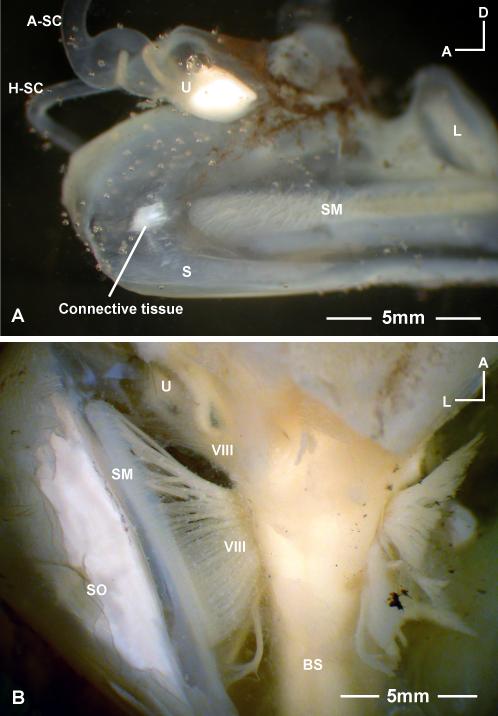
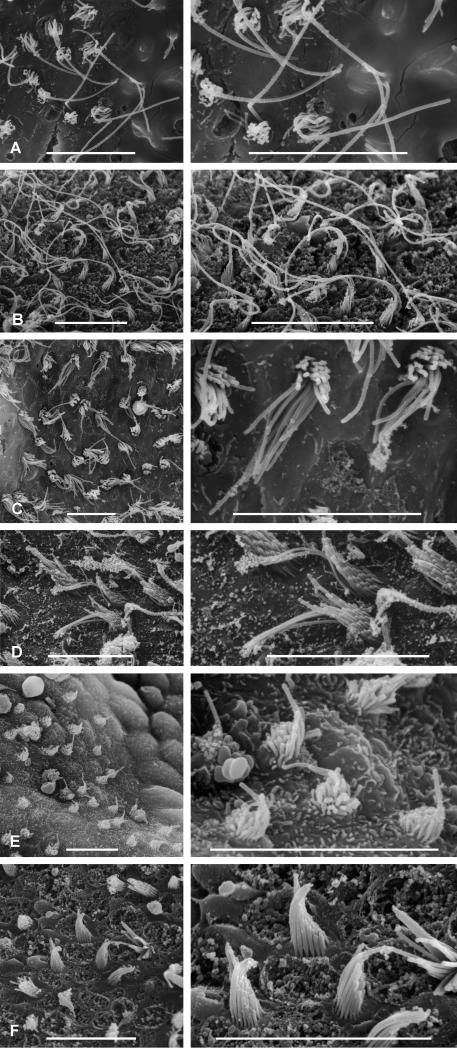
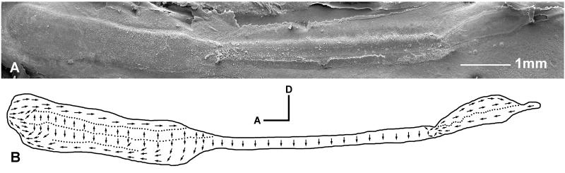
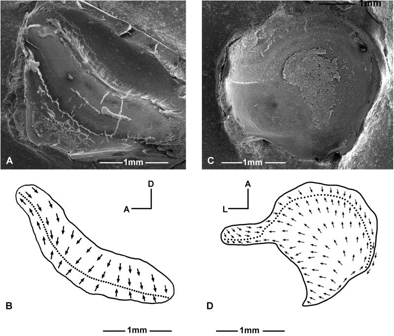
The inner ear structure of Antimora rostrata and its coupling to the swim bladder were analyzed and compared with the inner ears of several shallow-water species that also have similar coupling. The inner ear of Antimora has a long saccular otolith and sensory epithelium as compared to many other fishes. Some parts of the membranous labyrinth are thick and rigid, while other parts are thinner but attached tightly to the bony capsule. The partially rigid membranous labyrinth, along with its intimate connection to the swim bladder, may help the inner ear follow the sound oscillations from the swim bladder with better precision than would occur in a less rigid inner ear. In addition, the saccular sensory epithelium has an elaborate structure and an anterior enlargement that may be correlated with increased hearing sensitivity. Some of the features in the inner ear of Antimora may reflect the functional specialization of deep-water living and support the hypothesis that there is enhanced inner ear sensitivity in some deep-sea fishes.


Figures

Figure 1
Figure 1
Left and right inner ears of Antimora rostrata. The total length and standard length of the specimen are 440mm and 402mm, respectively. A: Medial view of a left inner ear showing the three end organs and their innervating eighth nerve branches. A measurement bar indicates the thickness of the posterior wall of the saccule. B: Lateral view of a right inner ear with all three otoliths clearly present in their pouches. A-SC, P-SC, H-SC, anterior, posterior, and horizontal semicircular canals; CC, common crux; L, lagena; LN, nerves to lagena; S, saccule; SN, nerves to saccule; SO, saccular otolith (sagitta). U, utricle; UN, nerves to utricle.
Figure 2
Figure 2
Lagena of Antimora rostrata. A: Lateral view of a right lagena. The walls of the sac are from 0.4 to 0.8mm thick. B: Left lagenar otolith and otolithic membrane taken out from the lagenar sac after osmium fixation. C: Medial view of the same otolith. The brown veil-like substance is the otolithic membrane (OM) stained by osmium. Note that the otolithic membrane extends out from two ends of the lagenar otolith. Dissections show that the otolithic membrane covers the regions of the sensory epithelium that are not covered by the otolith itself. LM, lagenar macula; OM, otolithic membrane.
Figure 3
Figure 3
Utricle of Antimora rostrata. A: Dorsal view of a right utricle with the otolith inside the pouch and the ampullae of the anterior (A-Amp) and horizontal semicircular canals (H-Amp) still attached. B: The utricular epithelium isolated from A, note that the striola (Str, more opaque yellowish region) and lacinia (Lac) are not covered by the otolith. C: Ventral view of the otolith taken from A, revealing the bumpy surface that is connected to the otolith membrane that lies between the otolith and the sensory epithelium. D: Side view of the same otolith to show the dome shape. UO, utricular otolith; UM, utricular macula.
Figure 4
Figure 4
Inner ears, brain, and swim bladder of Antimora rostrata (anterior to the top for A, B, and C). A: Dorsal view of the brain and inner ears after removing part of the skull and cartilage. B: Ventral view of the inner ears and the brainstem after removing part of the bottom of the cranium, with a clear indication of the rigidity of the saccule without the support of water. After removing the two anterior chambers of the swim bladder from where they attach to the inner ear’s bony capsules, the stubs of the dense connective tissue (indicated by white circles) indicate the attachment points. C: The relationship between the brain, inner ears, and swim bladder in Antimora; note that the size of the brain is relatively small compared with the size of the inner ears. D: Lateral view of the relative position between the inner ear and the swim bladder with respect to the head and the body. Also shown in D are two vertebrae (V) and a muscle bundle to the upper pharyngeal teeth (PT). AC, PC, anterior and posterior chamber of swim bladder; A-SC, H-SC, P-SC, anterior, horizontal, and posterior semicircular canals; BR, brain; L, lagena; LO, lagenar otolith; PT, pharyngeal teeth; S, saccule; SN, nerve to saccule; SO, saccular otolith; CT, connective tissue; U, utricle; UO, utricular otolith.
Figure 5
Figure 5
A: The bony capsule connection on the medial wall of a saccule in Antimora rostrata. This is a medial view of part of a right saccule. The contact is located just anterior to the saccular macula (SM) and is connected to the interior wall of the cranial bone via a connective tissue. B: Ventral view of the brainstem (BS) and right inner ear of Antimora, showing the array of auditory nerve fibers (VIII, eighth cranial nerve) spreading along either side of the brain stem. Refer to Figure 4 for other abbreviations.
Figure 6
Figure 6
Examples of hair bundles from the end organs of Antimora rostrata. A: Bundles from the ventral edge of the lagena (Fig. 8B). B: Bundles from the tip of lacinia on the utricle (Fig. 8D). C: Bundles from the striola of the lagena (Fig. 8B). D: Bundles from the caudal segment of the saccule (Fig. 7B). E: Bundles from the ventral tip of the utricle (Fig. 8D). F: Bundles from the center of the striola in the utricle (Fig. 8D). Scale bars = 10 μm.
Figure 7
Figure 7
Ultrastructure of the saccular sensory epithelium in Antimora rostrata. A: SEM photo of a left saccular macula. B: Hair bundle orientation pattern of the saccule. Note that the macula has three distinct segments with eight different bundle orientation groups.
Figure 8
Figure 8
Structure of the sensory epithelia of the lagena (A-B) and utricle (C-D) in Antimora rostrata. A and C: SEM photo of the left lagenar macula and left utricular macula. B and D: Hair bundle orientation pattern of the lagena and utricle.

Similar articles

Cited by

References

    1. Bailey DM, Bagley PM, Jamieson AJ, Collins MA, Priede IG. In situ investigation of burst swimming and muscle performance in the deep-sea fish Antimora rostrata. J. Exp. Mar. Biol. Ecol. 2003;286:295–311.
    1. Bierbaum G. Untersuchungen über den Bau der Gehörorgane von Tiefseefischen. Z. Wiss. Zool. 1914;III:281–380.
    1. Bregman AS. Auditory Scene Analysis: The Perceptual Organization of Sound. MIT Press; Cambridge, MA: 1990.
    1. Bregman AS, Peter Dallos, Donata Oertel. Auditory scene analysis. In: Basbaum AI, Kanenko A, Shepherd GM, Westheimer G, editors. The Senses: A Comprehensive Reference. Vol 3, Audition. Academic Press; San Diego: 2008. pp. 861–870.
    1. Buran BN, Deng X, Popper AN. Structural variation in the inner ears of four deep-sea Elopomorph fishes. J. Morph. 2005;265:215–225. - PubMed

LinkOut - more resources