. 2011 Apr 21;6(4):e18826.
doi: 10.1371/journal.pone.0018826.

Somatic mutagenesis with a Sleeping Beauty transposon system leads to solid tumor formation in zebrafish

Affiliations

Somatic mutagenesis with a Sleeping Beauty transposon system leads to solid tumor formation in zebrafish

Maura McGrail et al. PLoS One. .

Abstract

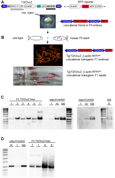
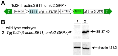
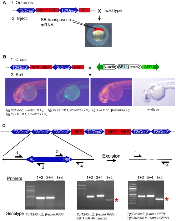
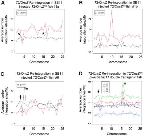
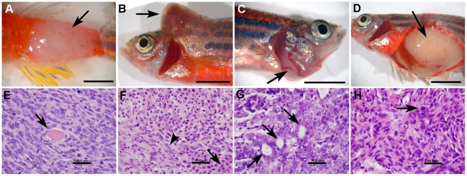
Large-scale sequencing of human cancer genomes and mouse transposon-induced tumors has identified a vast number of genes mutated in different cancers. One of the outstanding challenges in this field is to determine which genes, when mutated, contribute to cellular transformation and tumor progression. To identify new and conserved genes that drive tumorigenesis we have developed a novel cancer model in a distantly related vertebrate species, the zebrafish, Danio rerio. The Sleeping Beauty (SB) T2/Onc transposon system was adapted for somatic mutagenesis in zebrafish. The carp ß-actin promoter was cloned into T2/Onc to create T2/OncZ. Two transgenic zebrafish lines that contain large concatemers of T2/OncZ were isolated by injection of linear DNA into the zebrafish embryo. The T2/OncZ transposons were mobilized throughout the zebrafish genome from the transgene array by injecting SB11 transposase RNA at the 1-cell stage. Alternatively, the T2/OncZ zebrafish were crossed to a transgenic line that constitutively expresses SB11 transposase. T2/OncZ transposon integration sites were cloned by ligation-mediated PCR and sequenced on a Genome Analyzer II. Between 700-6800 unique integration events in individual fish were mapped to the zebrafish genome. The data show that introduction of transposase by transgene expression or RNA injection results in an even distribution of transposon re-integration events across the zebrafish genome. SB11 mRNA injection resulted in neoplasms in 10% of adult fish at ∼10 months of age. T2/OncZ-induced zebrafish tumors contain many mutated genes in common with human and mouse cancer genes. These analyses validate our mutagenesis approach and provide additional support for the involvement of these genes in human cancers. The zebrafish T2/OncZ cancer model will be useful for identifying novel and conserved genetic drivers of human cancers.


Conflict of interest statement

Competing Interests: The authors have declared that no competing interests exist.

Figures

Figure 1
Figure 1. Isolation of transgenic T2/OncZ concatemer lines.
(A) T2/OncZ transposon vector and RFP reporter used to isolate concatemers. IRL and IRR, left and right transposon inverted repeats; SA, splice acceptor from intron 1 of ß-actin gene; MSCV 5′ LTR, murine stem cell virus 5′ long terminal repeat; ß-actin, ß-actin promoter minus splice acceptor at 3′ end; SD, splice donor; En2-SA, splice acceptor from mouse engrailed 2 gene; pA, SV40 polyadenylation sequence; AFP, ocean pout antifreeze protein 3′ UTR; black bar represents probe used on genomic Southerns shown in panel C. Grey box represents probe used on genomic Southerns shown in panel D. Linear DNA fragments of T2/OncZ and the ß-actin:RFP reporter gene were mixed and co-injected into 1-cell zebrafish embryos. (B) Adult F0 founders were outcrossed to wild type and transgenic F1 embryos identified by ubiquitious RFP fluorescence. (C) Genomic Southern blots to estimate transposon copy number in Tg(T2/OncZ, ß-actin:RFP) concatemer lines 1–7. DNA was isolated from F2 generation heterozygous adults. (D) Genomic Southern blot of DNA isolated from F3 generation heterozygous Tg(T2/OncZ, ß-actin:RFP)is7 and Tg(T2/OncZ, ß-actin:RFP)is6 adults. Plasmid pT2/OncZ was loaded as reference in copy # control lanes.
Figure 2
Figure 2. Isolation of transgenic zebrafish expressing constitutive SB11 transposase.
(A) Diagram of miniTol2 vector containing a constitutive ß-actin promoter: SB11 cDNA cassette. 5′ and 3′, Tol2 inverted terminal repeats; zf ß-a 3′UTR, zebrafish ß-actin 3′ UTR; cmlc2, zebrafish cardiac myosin light chain 2 promoter. (B) Western blot demonstrates the expression of SB11 in Tg(Tol2<ß-actin:SB11, cmlc2:GFP>) transgenic embryos. The blot was stripped and re-probed with an anti-ß-actin antibody for loading control.
Figure 3
Figure 3. Two strategies for T2/OncZ insertional mutagenesis in zebrafish somatic tissues.
(A) Methods 1: Injection of in vitro transcribed SB11 mRNA at the 1-cell stage. (B) Method 2: Genetic cross between Tg(T2/OncZ, ß-actin:RFP) (T2/OncZ) and constitutive Tg(Tol2<ß-actin:SB11, cmlc2:GFP>) (ß-actin:SB11) fish. At 24 hpf embryos are sorted into 4 progeny classes. (C) Excision PCR assay on 5 dpf larvae to demonstrate mobilization of transposons out of the concatemer in the presence of SB11 transposase. Primers 1 and 4 amplify a 220 bp band (red asterisk) flanking the transposon excision site in the concatemer. Left panel, Tg(T2/OncZ, ß-actin:RFP)is6 larvae; middle panel, SB11 injected Tg(T2/OncZ, ß-actin:RFP)is6 larvae; right panel, double transgenic Tg(T2/OncZ, ß-actin:RFP)is6; Tg(Tol2<ß-actin:SB11, cmlc2:GFP>) larvae.
Figure 4
Figure 4. Re-integration of T2/OncZ after transient or constitutive SB11 transposase expression.
(A, B, C) The average number of re-integration sites per Mb plotted across all 25 chromosomes for tumor (T) and control (C) muscle samples from fish #1a, #1b and #6. Transposase source was supplied as SB11 mRNA injected at the 1-cell stage into Tg(T2/OncZ, ß-actin:RFP)is6 (fish #1a, #1b) or Tg(T2/OncZ, ß-actin:RFP)is7 (fish #6) embryos. (D) The average number of re-integration sites per Mb plotted across all 25 chromosomes for control samples from fish 1C, 2C, 3C, and 5C, and tumor samples from fish 2T and 8T. Fish were double heterozygous Tg(T2/OncZ, ß-actin:RFP)is6 ;Tg(Tol2<ß-actin:SB11, cmlc2:GFP>).
Figure 5
Figure 5. Histopathologic features of solid tumors in T2/OncZ mutagenized fish.
(A–D) gross images of neoplasms in fish 1a, 2, 6, and 8 respectively. (E–H) Histopathology of zebrafish neoplasms: Hematoxylin and Eosin stained sections at 1000× magnification. (E) Spindle cell sarcoma from fish 1a, note entrapped skeletal muscle fibers (arrow). (F) Mixed neoplasm from fish 2, neoplastic round cells (arrow) were intermixed with neoplastic spindle shaped cells (arrow head). (G) Carcinoma from fish 6, neoplastic cells were arranged into multiple acinar structures (arrow). (H) Spindle cell sarcoma from fish 8, note the mitotic figure (arrow). Scale bar A–D, 0.5 cm; E–H, 50 um.

Similar articles

Cited by

References

    1. Negrini S, Gorgoulis VG, Halazonetis TD. Genomic instability–an evolving hallmark of cancer. Nat Rev Mol Cell Biol. 2010;11:220–228. - PubMed
    1. Lengauer C, Kinzler KW, Vogelstein B. Genetic instability in colorectal cancers. Nature. 1997;386:623–627. - PubMed
    1. Campbell PJ, Yachida S, Mudie LJ, Stephens PJ, Pleasance ED, et al. The patterns and dynamics of genomic instability in metastatic pancreatic cancer. Nature. 2010;467:1109–1113. - PMC - PubMed
    1. Sjoblom T, Jones S, Wood LD, Parsons DW, Lin J, et al. The consensus coding sequences of human breast and colorectal cancers. Science. 2006;314:268–274. - PubMed
    1. Wood LD, Parsons DW, Jones S, Lin J, Sjoblom T, et al. The genomic landscapes of human breast and colorectal cancers. Science. 2007;318:1108–1113. - PubMed

Publication types