. 2011 Apr 21;6(4):e19090.
doi: 10.1371/journal.pone.0019090.

Loss of ATF2 function leads to cranial motoneuron degeneration during embryonic mouse development

Affiliations

Loss of ATF2 function leads to cranial motoneuron degeneration during embryonic mouse development

Julien Ackermann et al. PLoS One. .

Abstract

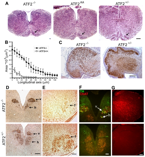
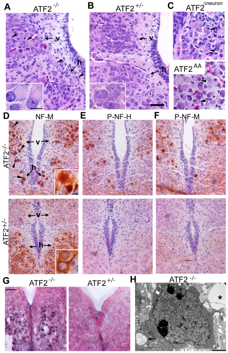
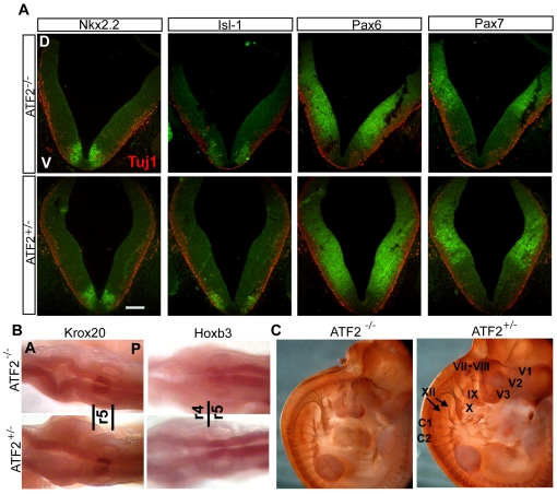
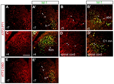
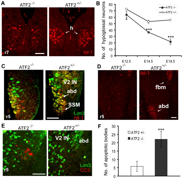
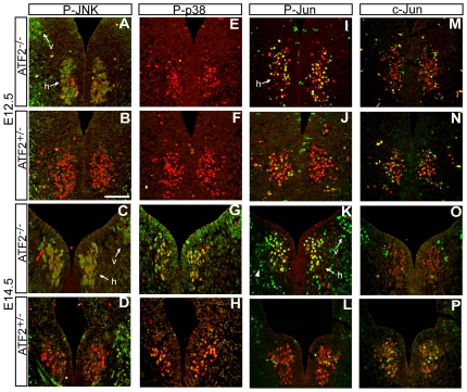
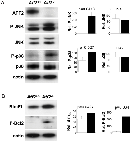
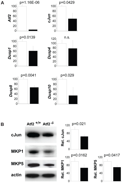
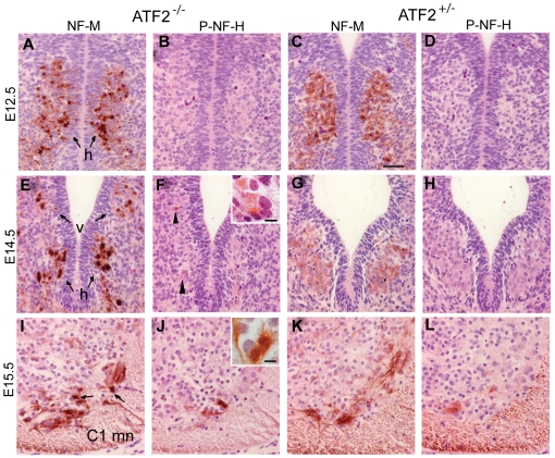
The AP-1 family transcription factor ATF2 is essential for development and tissue maintenance in mammals. In particular, ATF2 is highly expressed and activated in the brain and previous studies using mouse knockouts have confirmed its requirement in the cerebellum as well as in vestibular sense organs. Here we present the analysis of the requirement for ATF2 in CNS development in mouse embryos, specifically in the brainstem. We discovered that neuron-specific inactivation of ATF2 leads to significant loss of motoneurons of the hypoglossal, abducens and facial nuclei. While the generation of ATF2 mutant motoneurons appears normal during early development, they undergo caspase-dependent and independent cell death during later embryonic and foetal stages. The loss of these motoneurons correlates with increased levels of stress activated MAP kinases, JNK and p38, as well as aberrant accumulation of phosphorylated neurofilament proteins, NF-H and NF-M, known substrates for these kinases. This, together with other neuropathological phenotypes, including aberrant vacuolisation and lipid accumulation, indicates that deficiency in ATF2 leads to neurodegeneration of subsets of somatic and visceral motoneurons of the brainstem. It also confirms that ATF2 has a critical role in limiting the activities of stress kinases JNK and p38 which are potent inducers of cell death in the CNS.


Conflict of interest statement

Competing Interests: The authors have declared that no competing interests exist.

Figures

Figure 1
Figure 1. Histological abnormalities in E18.5 ATF2 mutant cerebellum and brainstem.
(A) Hematoxylin and eosin (H&E) stained transversal sections of brainstem at the level of the inferior olive. Atf2−/− and Atf2AA brainstems are significantly smaller and have an enlarged central canal compared to Atf2+/−. The inferior olive (arrow) is severely underdeveloped in mutant embryos. Bar: 250 µm. (B) Area plot of the central canal (mean ± SEM of 3 brainstems analysed per genotype), measured along the antero-posterior axis of serial transversal sections from the obex (0 µm on the x-axis) posteriorly to the caudal end of the medulla, shows a significant enlargement of the canal in Atf2−/− embryos. (C) Horseradish peroxidase (HRP) immunostaining of calbindin in sagittal sections of the cerebellum. Atf2−/− cerebellum lacks the foliation and the tripartite layering seen in Atf2+/− cerebellum. Bar: 250 µm. (D) HRP immunostaining of choline acetyltransferase (ChAT) in transversal sections of Atf2−/− and Atf2+/− posterior medulla. Number of hypoglossal motoneurons (h) is bilaterally decreased in Atf2−/− embryos while number of dorsal vagal motoneurons (v) appears normal. This was seen at several levels along the longitudinal axis. Bar: 200 µm. (E) HRP immunostaining of Islet-1 (Isl-1) in transversal sections of Atf2−/− and Atf2+/− anterior medulla. A severe reduction in the number of motoneurons is found in the Atf2−/− facial nucleus (f). Bar: 100 µm. (F) Double immunofluorescence staining of TH (green) and ChAT (red) shows aberrant expression of TH in hypoglossal (h) and dorsal vagal (v) motoneurons in Atf2−/− brains. Bar: 50 µm. (G) GFAP immunostaining (red) reveals aberrant expression of GFAP in the mantle zone of Atf2−/− brainstem. Bar: 50 µm.
Figure 2
Figure 2. Neuropathological lesions in E18.5 ATF2 mutant motoneurons.
(A, B) H&E stained transversal sections of Atf2−/− and Atf2+/− posterior medulla. Atf2−/− hypoglossal (h) and dorsal vagal (v) motoneurons show ballooned perikarya with eccentrically positioned nuclei (arrowheads). Insets: magnification of dorsal vagal motoneurons. (C) H&E staining of Atf2Δneuron hypoglossal and Atf2AA vagal motoneurons showing the same pathological lesions (arrowheads). (D–F) HRP immunostaining with antibodies against neurofilament M (NF-M), phospho-neurofilament H (P-NF-H, RMO24.9) and phospho-neurofilament M (P-NF-M, RMO8) in Atf2−/− and Atf2+/− posterior medulla. Aberrant NF-M accumulation in the soma of Atf2−/− dorsal vagal and hypoglossal motoneurons is indicated by arrowheads. P-NF-H and P-NF-M are predominantly present in Atf2−/− motoneurons. Insets: magnification of NF-M stained hypoglossal motoneurons. (G) Sudan black B staining shows lipid accumulation in Atf2−/− but not in Atf2+/− hypoglossal motoneurons. Bars: A–G, 100 µm; insets, 25 µm.(H) Transmission electron microscopy photograph of Atf2−/− brainstem shows a lipid droplet near a cell (asterisk), and a cytoplasmic vacuole filled with neurofibrillary material (arrow). Bar: 2 µm.
Figure 3
Figure 3. Dorso-ventral (D–V) and antero-posterior (A–P) patterning is normal in Atf2 / brainstem at E10.5.
(A) Double immunofluorescence staining for class III β-tubulin (Tuj1, red) and for neuronal class progenitor cells markers Nkx2.2, Pax6, Pax7 (all green) and postmitotic marker Isl-1 (green) in Atf2−/− and Atf2+/− brainstem. No significant difference was observed in the expression domains of the transcription factors between the two genotypes. Bar: 100 µm. (B) Whole-mount in situ hybridization for Krox20 and Hoxb3. No differences were observed in the expression domain between the two genotypes. r4, r5, rhombomere 4 and 5. Note that, at this stage, Krox20 expression is seen in rhombomere 5 (r5) only. (C) Atf2−/− and Atf2+/− whole-mount immunostaining with NF-M antibody at E12.5. Fiber nerves extend normally from Atf2−/− hypoglossal (XII) and vagal (X) nuclei. C1, C2, first and second cervical nerve, XII, hypoglossal nerve, X, IX, vagal and glossopharyngeal nerves, VII, VIII, acousticofacial nerve, V3, mandibular nerve, V2, maxillary nerve, V1, ophthalmic nerve.
Figure 4
Figure 4. ATF2 expression in somatic and visceral motoneurons at E11.5 and E12.5.
(A–D) Fluorescence immunostaining for phosphorylated ATF2 (ATF2-PT71, red). (A'–D') Double immunofluorescence staining for phosphorylated ATF2 (red) and postmitotic marker Isl-1 (green). Phosphorylated ATF2 is robustly detected in E12.5 hypoglossal (h) and dorsal vagal (v) motoneurons (A, A'), in E12.5 abducens (abd) motoneurons (B, B', arrowheads) and in facial branchiomotor neurons (C, C') at E11.5. In addition phospho-ATF2 is also detected in C1 motoneurons of the spinal cord at E13.5 (D, D', arrowheads). Little or no expression of phosphorylated ATF2 is found in the surrounding cells or on the dorsal side of the brainstem at this stage. Bar: 100 µm. (E, E') E12.5 hypoglossal (h) and dorsal vagal (v) motoneurons express ATF2 as shown by immunostaining with ATF2-DBD antibody (red) and Isl-1 (green). r4, r5, r7, rhombomere, 4, 5, and 7.
Figure 5
Figure 5. Somatic motoneuron defects in Atf2 / embryos.
(A) Isl-1 immunostaining (red) of Atf2−/− and Atf2+/− hypoglossal motoneurons at E14.5 reveal the reduction of Isl-1 positive hypoglossal motoneurons in Atf2−/− embryos. (B) Quantitative analysis of hypoglossal motoneurons (mean number per section ± SD) in serial transversal sections of rhombomere 7 (r7) by Isl-1 immunostaining for E12.5 and E14.5 embryos or by 5-HT immunostaining and hematoxylin counterstaining for E18.5 embryos. Student's two-tailed t-Test, *, p = 0.04; ***, p<0.01; ***; p<0.001. (C) Coimmunostaining of abducens motoneurons with Isl-1 (red) and Lim3 (green) antibodies reveals normal production of these motoneurons in Atf2−/− mice at E10.5 in r5. V2 IN, V2 interneurons, abd, abducens motoneurons, SSM, superior salivatory motoneurons. (D) Complete loss of abducens motoneurons (abd) in Atf2−/− E12.5 embryos as revealed by Isl-1 (red) immunostaining. fbm, facial branchiomotor neurons. (E) Coimmunostaining of abducens motoneurons with Cleaved Caspase 3 (CC3, red) and Lim3 (green) antibodies reveals increased apoptosis in Atf2−/− motoneurons at E11.5. Lim3 strongly labels V2 interneurons and more weakly abducens motoneurons. Bar: 100 µm. (F) Quantitative analysis of apoptotic bodies (mean number per section ± SD) in serial transversal sections of rhombomere 5 at E11.5 reveals increased apoptosis of abducens neurons in Atf2−/− embryos. ***, p<0.01.
Figure 6
Figure 6. Enhanced phosphorylation of JNK, p38, and c-Jun in Atf2 / motoneurons.
Double immunofluorescence staining of Isl-1 (A–P, red) and phospho-JNK (Thr183/Tyr185, green, A–D), phospho-p38 (Thr180/Tyr182, green, E-H), phospho-c-Jun (Ser73, green, I-L) and pan-c-Jun (green, M–P) in hypoglossal (h) and vagal (v) motoneurons at E12.5 and E14.5. (A–D) JNK is hyperphosphorylated in both motoneuron types in Atf2−/− (A and C) compared to Atf2+/− (B and D) embryos at E12.5 and E14.5. (E–H) Phosphorylation of p38 is hardly detectable in Atf2+/− and Atf2−/− at E12.5 (E and F) but significantly increases at E14.5 in motoneurons of Atf2−/− (G) compared to Atf2+/− embryos (H). (I-L) c-Jun is hyperphosphorylated in motoneurons of Atf2−/− (I and K) compared to Atf2+/− (J and L) embryos at E12.5 and E14.5. Strong phosphorylation of c-Jun is notable in hypoglossal and vagal motoneurons at E14.5 and there is a weaker but detectable phosphorylation signal in surrounding cells in Atf2−/− embryos (K, arrowheads). (M, N) High and moderate levels of c-Jun were observed in hypoglossal motoneurons but were indistinguishable between Atf2−/− and Atf2+/− embryos at E12.5. (O, P) At E14.5, Atf2−/− hypoglossal neurons tend to express c-Jun at lower levels than in Atf2+/− neurons; notably some strong c-Jun labeling was detected in some Isl-1 negative cells in Atf2−/−. Bar: 100 µm.
Figure 7
Figure 7. Up-regulation of P-JNK and P-p38 in Atf2 / hindbrains.
(A) Western blot analysis shows increased P-JNK (Thr183/Tyr185) and P-p38 (Thr180/Tyr182) in Atf2−/− compared to Atf2+/+ E14.5 hindbrains extracts. (B) Expression levels of BimEL and P-Bcl2 (Ser70) are shown. Bar graphs show relative signal intensity between Atf2+/+ (white bars) compared to Atf2−/− (black bars). Significance values (p) were determined from 3 independent samples using Student's t-test.
Figure 8
Figure 8. Expression of ATF2 target genes in hindbrain motoneurons.
Real-time quantitative PCR assays of laser micro-dissected tissue of E14.5 hindbrains. Bar graphs show relative expression values between Atf2+/+ (white bars) and Atf2−/− (black bars) for Atf2, c-Jun, Dusp1, Dusp4, Dusp8, and Dusp10 mRNAs. Significance values (p) were determined from 3 independent samples using Student's t-test.
Figure 9
Figure 9. Neurofilament M (NF-M) accumulation in Atf2 / motoneurons.
(A–D) Immunostaining against NF-M (A and C) and phosphorylated NF-H (B and D) at E12.5 reveals strong accumulation of NF-M in the soma of Atf2−/− hypoglossal motoneurons (h) but not in Atf2+/− motoneurons (compare A and C). Phosphorylation of NF-H as detected with the antibody RMO24.9 was not yet observed at this age. (E–H) Immunostaining against NF-M (E and G) and phosphorylated NF-H (F and H) at E14.5 reveals strong accumulation of NF-M in hypoglossal (h) and vagal (v) motoneurons in Atf2−/− (E) compared to Atf2+/− embryos (G). Phosphorylation of NF-H can be detected in Atf2−/− motoneurons (F, arrowheads) but not in Atf2+/− neurons (H). Inset: magnification of P-NF-H stained hypoglossal neurons. (I-L) Immunostaining against NF-M (I and K) and phosphorylated NF-H (J and L) at E15.5 reveals aberrant accumulation of NF-M and hyperphosphorylation of NF-H in the soma of Atf2−/− spinal cord motoneurons at the C1 level (C1 mn) but not in Atf2+/− neurons. Bar: 100 µm. Inset: magnification of P-NF-H stained C1 motoneurons. Bar: 25 µm.

Similar articles

Cited by

References

    1. Herdegen T, Leah JD. Inducible and constitutive transcription factors in the mammalian nervous system: control of gene expression by Jun, Fos and Krox, and CREB/ATF proteins. Brain Res Brain Res Rev. 1998;28:370–490. - PubMed
    1. Gupta S, Campbell D, Derijard B, Davis RJ. Transcription factor ATF2 regulation by the JNK signal transduction pathway. Science. 1995;267:389–393. - PubMed
    1. Ouwens DM, de Ruiter ND, van der Zon GC, Carter AP, Schouten J, et al. Growth factors can activate ATF2 via a two-step mechanism: phosphorylation of Thr71 through the Ras-MEK-ERK pathway and of Thr69 through RalGDS-Src-p38. Embo J. 2002;21:3782–3793. - PMC - PubMed
    1. van Dam H, Wilhelm D, Herr I, Steffen A, Herrlich P, et al. ATF-2 is preferentially activated by stress-activated protein kinases to mediate c-jun induction in response to genotoxic agents. Embo J. 1995;14:1798–1811. - PMC - PubMed
    1. Livingstone C, Patel G, Jones N. ATF-2 contains a phosphorylation-dependent transcriptional activation domain. Embo J. 1995;14:1785–1797. - PMC - PubMed

Publication types

MeSH terms

Substances