. 2011 Apr;7(4):e1002035.
doi: 10.1371/journal.pcbi.1002035. Epub 2011 Apr 21.

In silico elucidation of the recognition dynamics of ubiquitin

Affiliations

In silico elucidation of the recognition dynamics of ubiquitin

Dong Long et al. PLoS Comput Biol. 2011 Apr.

Abstract

Elucidation of the mechanism of biomacromolecular recognition events has been a topic of intense interest over the past century. The inherent dynamic nature of both protein and ligand molecules along with the continuous reshaping of the energy landscape during the binding process renders it difficult to characterize this process at atomic detail. Here, we investigate the recognition dynamics of ubiquitin via microsecond all-atom molecular dynamics simulation providing both thermodynamic and kinetic information. The high-level of consistency found with respect to experimental NMR data lends support to the accuracy of the in silico representation of the conformational substates and their interconversions of free ubiquitin. Using an energy-based reweighting approach, the statistical distribution of conformational states of ubiquitin is monitored as a function of the distance between ubiquitin and its binding partner Hrs-UIM. It is found that extensive and dense sampling of conformational space afforded by the µs MD trajectory is essential for the elucidation of the binding mechanism as is Boltzmann sampling, overcoming inherent limitations of sparsely sampled empirical ensembles. The results reveal a population redistribution mechanism that takes effect when the ligand is at intermediate range of 1-2 nm from ubiquitin. This mechanism, which may be depicted as a superposition of the conformational selection and induced fit mechanisms, also applies to other binding partners of ubiquitin, such as the GGA3 GAT domain.


Conflict of interest statement

The authors have declared that no competing interests exist.

Figures

Figure 1
Figure 1. Structural analysis of X-ray crystal structures, EROS ensemble, and MD ensemble.
(A) Cα root mean square fluctuation (RMSF) of MD ensemble (black), EROS ensemble (green), and 19 ubiquitin X-ray crystal structures (magenta). (B) Residuewise root mean square deviation (RMSD) values (Eq. (1)) for the pair-wise structural comparisons of X-ray crystal structures and their closest EROS conformers (green solid line) and X-ray crystal structures and their closest MD conformers (1 million structures) (black solid line). Green peaks that belong to protein binding regions are circled with dash-dot lines. (C) The Cα RMSD calculated for each X-ray crystal structure with the closest EROS conformer (x-axis) and the closest MD conformer (y-axis). The RMSD values of the MD conformers are substantially smaller than for the EROS conformers. Definitions of RMSF and RMSD parameters are given in Text S1.
Figure 2
Figure 2. Projection of the structural ensembles on 2D PCA space.
(A) Comparison of the X-ray structures (magenta diamonds), EROS ensemble (green triangles) and MD ensemble (black dots) in 2D space spanned by the 2 largest principal components. PCA was performed using the 19 X-ray crystal bound forms only. (B) Population distribution of MD ensemble on two dimensions, which can be grouped into the three substates S1, S2, and S3.
Figure 3
Figure 3. Autocorrelation function of the principal mode motion.
The autocorrelation function C(t) of the internal dynamics along the first principal component (Eq. S5; blue line) is fitted with a two-exponential function (Eq. S6; red line). The parameters extracted by curve fitting are: a = 0.49; τfast = 0.4 ns; τslow = 12.9 ns.
Figure 4
Figure 4. Conformational space accessible to ubiquitin in complex with Hrs-UIM.
(A) 2D projection of complex MD ensemble (black dots). The corresponding crystal conformations are indicated by the magenta diamond for chain A (PDB code: 2D3G) and the magenta ‘+’ symbol for chain B of the same PDB entry. (B) Pseudo color presentation of population distribution of the ubiquitin bound form.
Figure 5
Figure 5. Population redistribution of substates of ubiquitin upon the approach of Hrs-UIM.
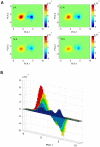
(A) The two-dimensional difference maps of the population densities were calculated in the presence of Hrs-UIM in the range from 9 to 18 Å away from its position in the crystal complex (PDB code 2D3G, chain P). (B) One-dimensional projection of the population difference maps along the largest principal component. Hrs-UIM was introduced at distances of 9 Å (yellow), 12 Å (green), 15 Å (cyan), and 18 Å (light blue). The ubiquitin populations in the free (dark blue) and bound (red) forms are included for comparison. For clarity, the populations of the free and bound states are scaled by 1/19 and 1/13 in the plot. It can be seen that the substate S3 becomes increasingly populated as Hrs-UIM approaches the binding site.
Figure 6
Figure 6. Schematic illustration of the population redistribution upon the approach of Hrs-UIM to the binding interface.
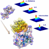
The population distribution at an intermediate distance (between 0 and 9 Å) is represented by the linear combination of the population distributions at 0 and 9 Å with weights of 30% and 70%, respectively. The four population density plots have the same axes as Figure 2B. The reshaping of the energy landscape of ubiquitin occurs in a continuous way, starting at nanometer distance range between the binding partners.

Similar articles

Cited by

References

    1. Fischer E. Einfluss der configuration auf die Wirkung der enzyme. Ber Dt Chem Ges. 1894;27:2985–2993.
    1. Koshland DE. Application of a theory of enzyme specificity to protein synthesis. Proc Natl Acad Sci USA. 1958;44:98–104. - PMC - PubMed
    1. Tsai CJ, Ma B, Nussinov R. Folding and binding cascades: shifts in energy landscapes. Proc Natl Acad Sci USA. 1999;96:9970–9972. - PMC - PubMed
    1. Ma B, Shatsky M, Wolfson HJ, Nussinov R. Multiple diverse ligands binding at a single protein site: a matter of pre-existing populations. Protein Sci. 2002;11:184–197. - PMC - PubMed
    1. Boehr DD, Nussinov R, Wright PE. The role of dynamic conformational ensembles in biomolecular recognition. Nat Chem Biol. 2009;5:789–796. - PMC - PubMed

Publication types