

Proline-rich tyrosine kinase 2 (Pyk2) promotes cell motility of hepatocellular carcinoma through induction of epithelial to mesenchymal transition
- PMID: 21533080
- PMCID: PMC3080371
- DOI: 10.1371/journal.pone.0018878
Proline-rich tyrosine kinase 2 (Pyk2) promotes cell motility of hepatocellular carcinoma through induction of epithelial to mesenchymal transition
Abstract
Aims: Proline-rich tyrosine kinase 2 (Pyk2), a non-receptor tyrosine kinase of the focal adhesion kinase (FAK) family, is up-regulated in more than 60% of the tumors of hepatocellular carcinoma (HCC) patients. Forced overexpression of Pyk2 can promote the proliferation and invasion of HCC cells. In this study, we aimed to explore the underlying molecular mechanism of Pyk2-mediated cell migration of HCC cells.
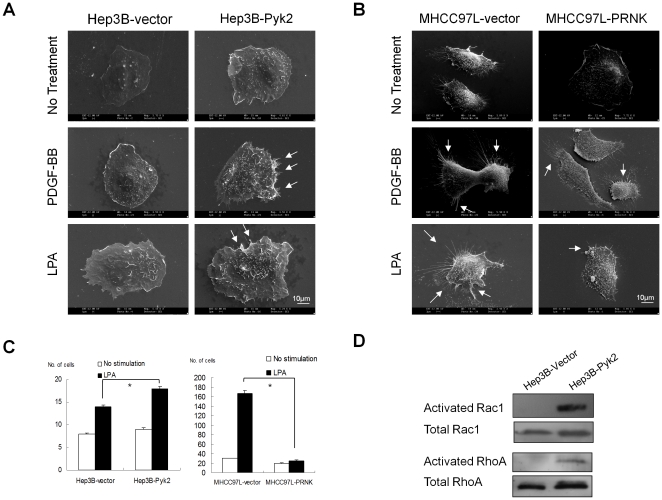
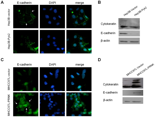
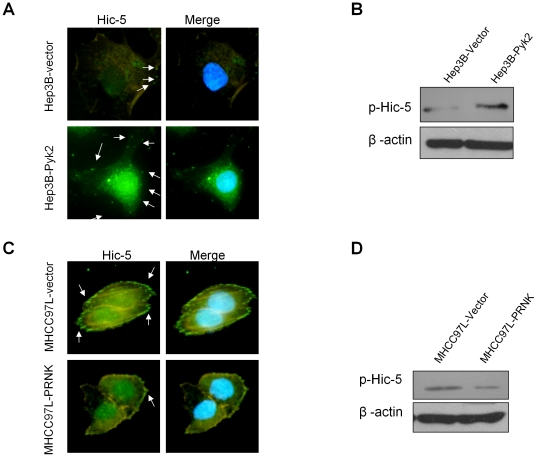
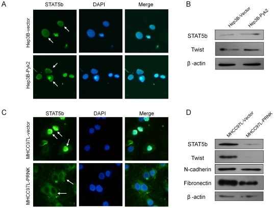
Methodology/principal findings: We demonstrated that Pyk2 transformed the epithelial HCC cell line Hep3B into a mesenchymal phenotype via the induction of epithelial to mesenchymal transition (EMT), signified by the up-regulation of membrane ruffle formation, activation of Rac/Rho GTPases, down-regulation of epithelial genes E-cadherin and cytokeratin as well as promotion of cell motility in presence of lysophosphatidic acid (LPA). Suppression of Pyk2 by overexpression of dominant negative PRNK domain in the metastatic HCC cell line MHCC97L transformed its fibroblastoid phenotype to an epithelial phenotype with up-regulation of epithelial genes, down-regulation of mesenchymal genes N-cadherin and STAT5b, and reduction of LPA-induced membrane ruffle formation and cell motility. Moreover, overexpression of Pyk2 in Hep3B cells promoted the phosphorylation and localization of mesenchymal gene Hic-5 onto cell membrane while suppression of Pyk2 in MHCC97L cells attenuated its phosphorylation and localization.
Conclusion: These data provided new evidence of the underlying mechanism of Pyk2 in controlling cell motility of HCC cells through regulation of genes associated with EMT.
Conflict of interest statement
Figures
References
-
- Farazi PA, DePinho RA. Hepatocellular carcinoma pathogenesis: from genes to environment. Nat Rev Cancer. 2006;6:674–687. - PubMed
-
- Mathurin P, Rixe O, Carbonell N, Bernard B, Cluzel P, et al. Review article: Overview of medical treatments in unresectable hepatocellular carcinoma–an impossible meta-analysis? Aliment Pharmacol Ther. 1998;12:111–126. - PubMed
-
- Kawano Y, Sasaki A, Kai S, Endo Y, Iwaki K, et al. Prognosis of patients with intrahepatic recurrence after hepatic resection for hepatocellular carcinoma: A retrospective study. Eur J Surg Oncol 2008 - PubMed
-
- Liotta LA. Mechanisms of cancer invasion and metastasis. Important Adv Oncol. 1985:28–41. - PubMed
-
- Stracke ML, Liotta LA. Multi-step cascade of tumor cell metastasis. In Vivo. 1992;6:309–316. - PubMed
Publication types
MeSH terms
Substances
LinkOut - more resources
Full Text Sources
Medical
Molecular Biology Databases
Research Materials
Miscellaneous
